6RMG image
Deposition Date 2019-05-06
Release Date 2019-10-09
Last Version Date 2024-11-20
Entry Detail
PDB ID:
6RMG
Title:
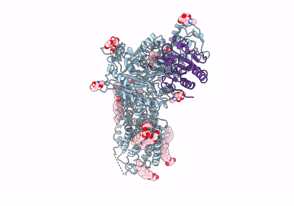
Structure of PTCH1 bound to a modified Hedgehog ligand ShhN-C24II
Biological Source:
Source Organism:
Homo sapiens (Taxon ID: 9606)
Eimeria acervulina (Taxon ID: 5801)
Host Organism:
Method Details:
Experimental Method:
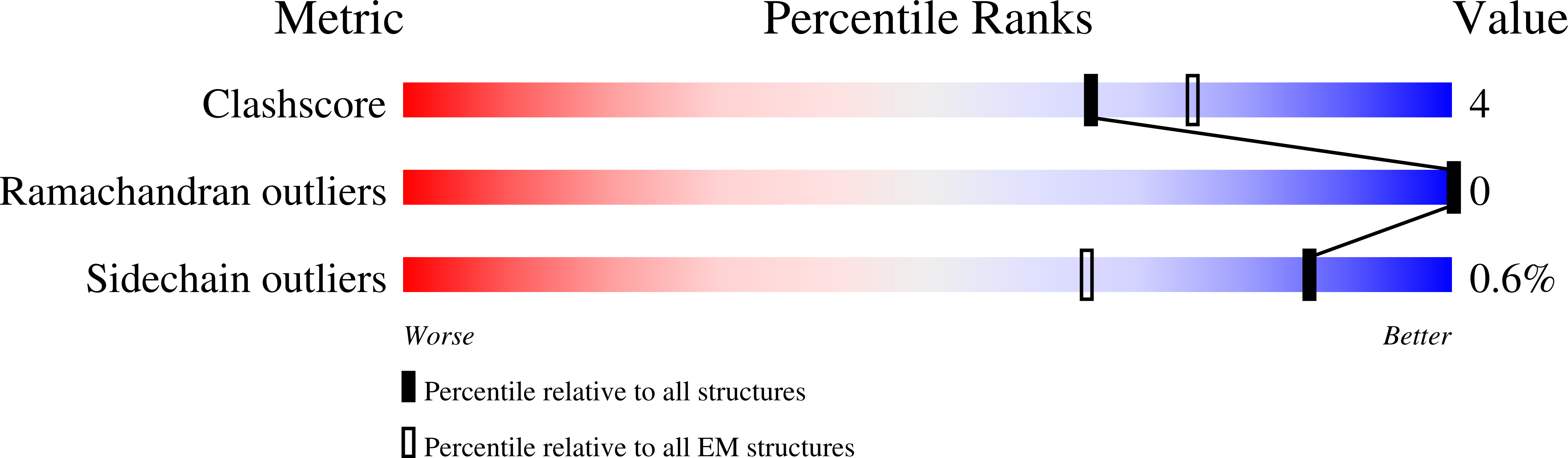
Resolution:
3.40 Å
Aggregation State:
PARTICLE
Reconstruction Method:
SINGLE PARTICLE
Macromolecular Entities
Protein Blast
Polymer Type:polypeptide(L)
Molecule:Protein patched homolog 1,GFP-like fluorescent chromoprotein FP506, related
Gene (Uniprot):PTCH1, EAH_00062270
Chain IDs:A
Chain Length:1471
Number of Molecules:1
Biological Source:Homo sapiens, Eimeria acervulina
Structures with similar UniProt ID
Protein Blast
Polymer Type:polypeptide(L)
Molecule:Sonic hedgehog protein
Gene (Uniprot):SHH
Chain IDs:B
Chain Length:285
Number of Molecules:1
Biological Source:Homo sapiens
Primary Citation
Structural basis of sterol recognition by human hedgehog receptor PTCH1.
Sci Adv 5 eaaw6490 eaaw6490 (2019)
PMID: 31555730 DOI: 10.1126/sciadv.aaw6490

Abstact

Hedgehog signaling is central in embryonic development and tissue regeneration. Disruption of the pathway is linked to genetic diseases and cancer. Binding of the secreted ligand, Sonic hedgehog (ShhN) to its receptor Patched (PTCH1) activates the signaling pathway. Here, we describe a 3.4-Å cryo-EM structure of the human PTCH1 bound to ShhNC24II, a modified hedgehog ligand mimicking its palmitoylated form. The membrane-embedded part of PTCH1 is surrounded by 10 sterol molecules at the inner and outer lipid bilayer portion of the protein. The annular sterols interact at multiple sites with both the sterol-sensing domain (SSD) and the SSD-like domain (SSDL), which are located on opposite sides of PTCH1. The structure reveals a possible route for sterol translocation across the lipid bilayer by PTCH1 and homologous transporters.

Legend

Protein

Chemical

Disease

Primary Citation of related structures