6RM9 image
Entry Detail
PDB ID:
6RM9
Keywords:
Title:
Crystal structure of the DEAH-box ATPase Prp2 in complex with Spp2 and ADP
Biological Source:
PDB Version:
Deposition Date:
2019-05-06
Release Date:
2020-02-05
Method Details:
Experimental Method:
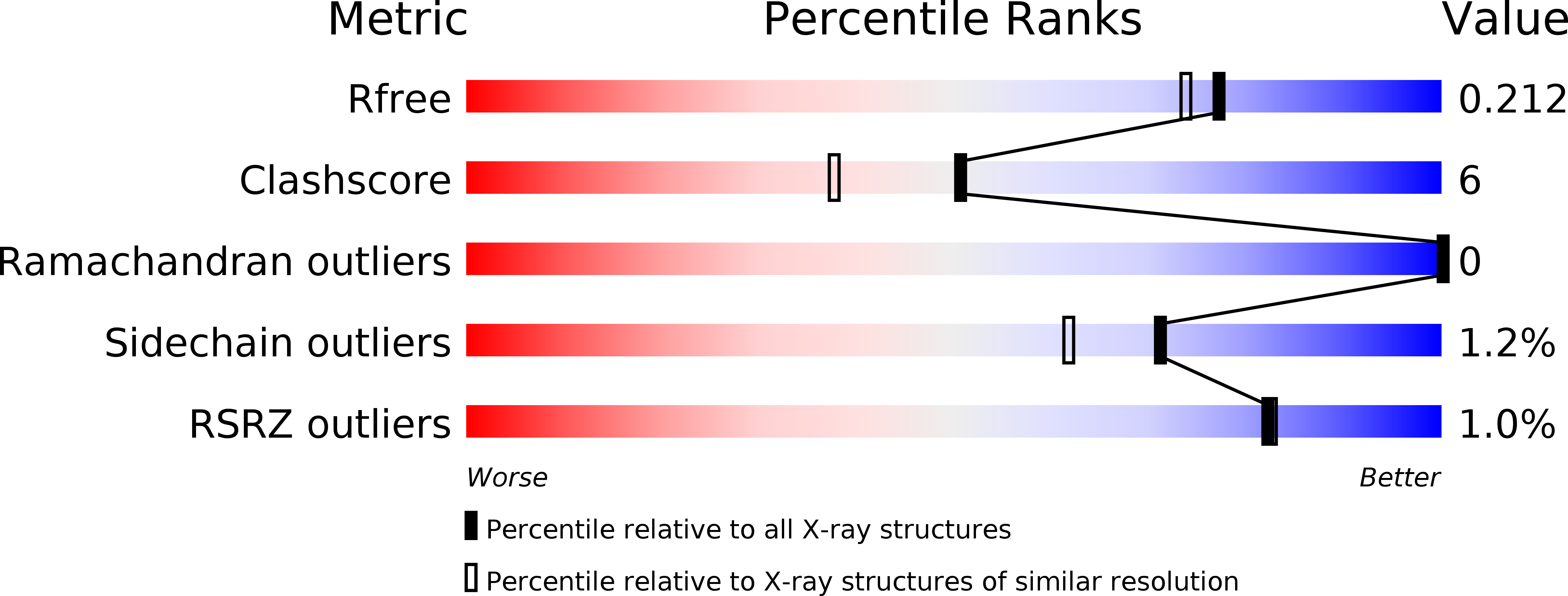
Resolution:
1.85 Å
R-Value Free:
0.21
R-Value Work:
0.19
R-Value Observed:
0.19
Space Group:
P 21 21 21
Macromolecular Entities
Polymer Type:polypeptide(L)
Description:Putative mRNA splicing factor
Chain IDs:A
Chain Length:660
Number of Molecules:1
Biological Source:Chaetomium thermophilum var. thermophilum DSM 1495
Polymer Type:polypeptide(L)
Description:Putative pre-mRNA splicing protein
Chain IDs:B (auth: C)
Chain Length:47
Number of Molecules:1
Biological Source:Chaetomium thermophilum var. thermophilum DSM 1495
Modified Residue
Compound ID Chain ID Parent Comp ID Details 2D Image
CSO A CYS modified residue
Primary Citation
Structural analysis of the intrinsically disordered splicing factor Spp2 and its binding to the DEAH-box ATPase Prp2.
Proc.Natl.Acad.Sci.USA 117 2948 2956 (2020)
PMID: 31974312 DOI: 10.1073/pnas.1907960117

Abstact

The spliceosome consists of five small RNAs and more than 100 proteins. Almost 50% of the human spliceosomal proteins were predicted to be intrinsically disordered or to contain disordered regions, among them the G-patch protein Spp2. The G-patch region of Spp2 binds to the DEAH-box ATPase Prp2, and both proteins together are essential for promoting the transition from the Bact to the catalytically active B* spliceosome. Here we show by circular dichroism and nuclear magnetic resonance (NMR) spectroscopy that Spp2 is intrinsically disordered in solution. Crystal structures of a complex consisting of Prp2-ADP and the G-patch domain of Spp2 demonstrate that the G-patch gains a defined fold when bound to Prp2. While the N-terminal region of the G-patch always folds into an α-helix in five different crystal structures, the C-terminal part is able to adopt two alternative conformations. NMR studies further revealed that the N-terminal part of the Spp2 G-patch, which is the most conserved region in different G-patch proteins, transiently samples helical conformations, possibly facilitating a conformational selection binding mechanism. The structural analysis unveils the role of conserved residues of the G-patch in the dynamic interaction mode of Spp2 with Prp2, which is vital to maintain the binding during the Prp2 domain movements needed for RNA translocation.

Legend

Protein

Chemical

Disease

Primary Citation of related structures