Deposition Date
1991-06-21
Release Date
1993-10-31
Last Version Date
2024-10-09
Entry Detail
PDB ID:
6RLX
Keywords:
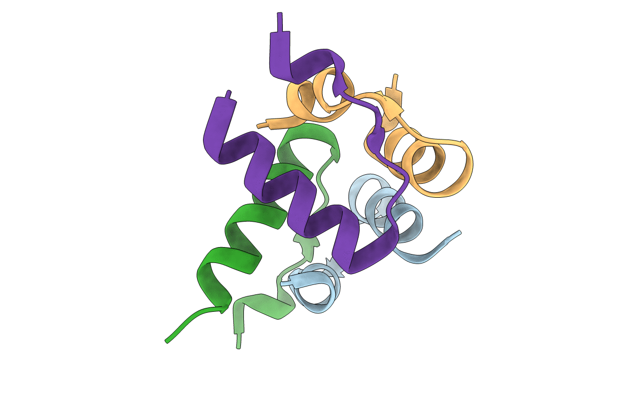
Title:
X-RAY STRUCTURE OF HUMAN RELAXIN AT 1.5 ANGSTROMS. COMPARISON TO INSULIN AND IMPLICATIONS FOR RECEPTOR BINDING DETERMINANTS
Biological Source:
Source Organism(s):
Homo sapiens (Taxon ID: 9606)
Expression System(s):
Method Details:
Experimental Method:
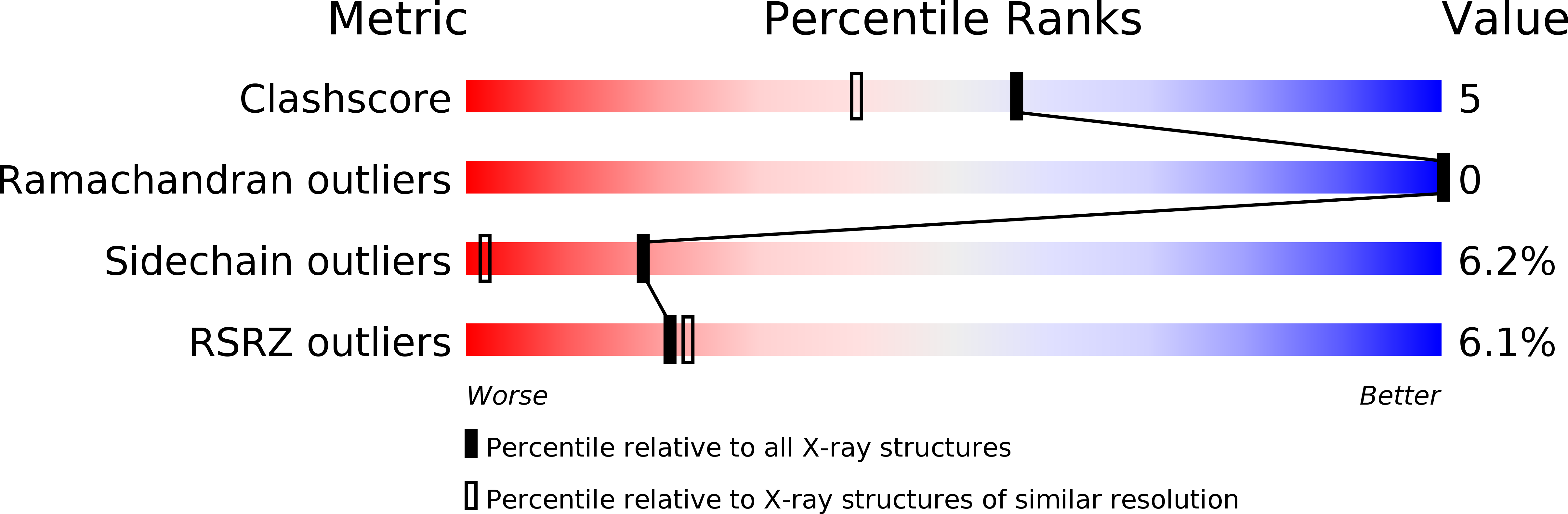
Resolution:
1.50 Å
R-Value Work:
0.18
R-Value Observed:
0.18
Space Group:
C 2 2 21