
Deposition Date
2019-04-26
Release Date
2019-10-16
Last Version Date
2024-01-24
Entry Detail
PDB ID:
6RJE
Keywords:
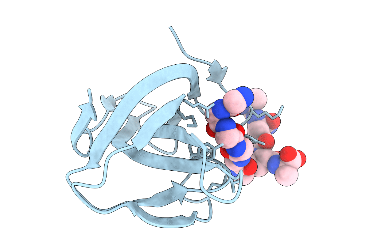
Title:
Lysostaphin SH3b P4-G5 complex, homesource dataset
Biological Source:
Source Organism(s):
Staphylococcus simulans (Taxon ID: 1286)
Expression System(s):
Method Details:
Experimental Method:
Resolution:
2.50 Å
R-Value Free:
0.29
R-Value Work:
0.25
R-Value Observed:
0.25
Space Group:
P 41 21 2
