Deposition Date
2019-04-04
Release Date
2019-12-25
Last Version Date
2024-01-24
Entry Detail
PDB ID:
6R9W
Keywords:
Title:
Crystal structure of InhA in complex with AP-124 inhibitor
Biological Source:
Source Organism(s):
Mycobacterium tuberculosis H37Rv (Taxon ID: 83332)
Expression System(s):
Method Details:
Experimental Method:
Resolution:
1.75 Å
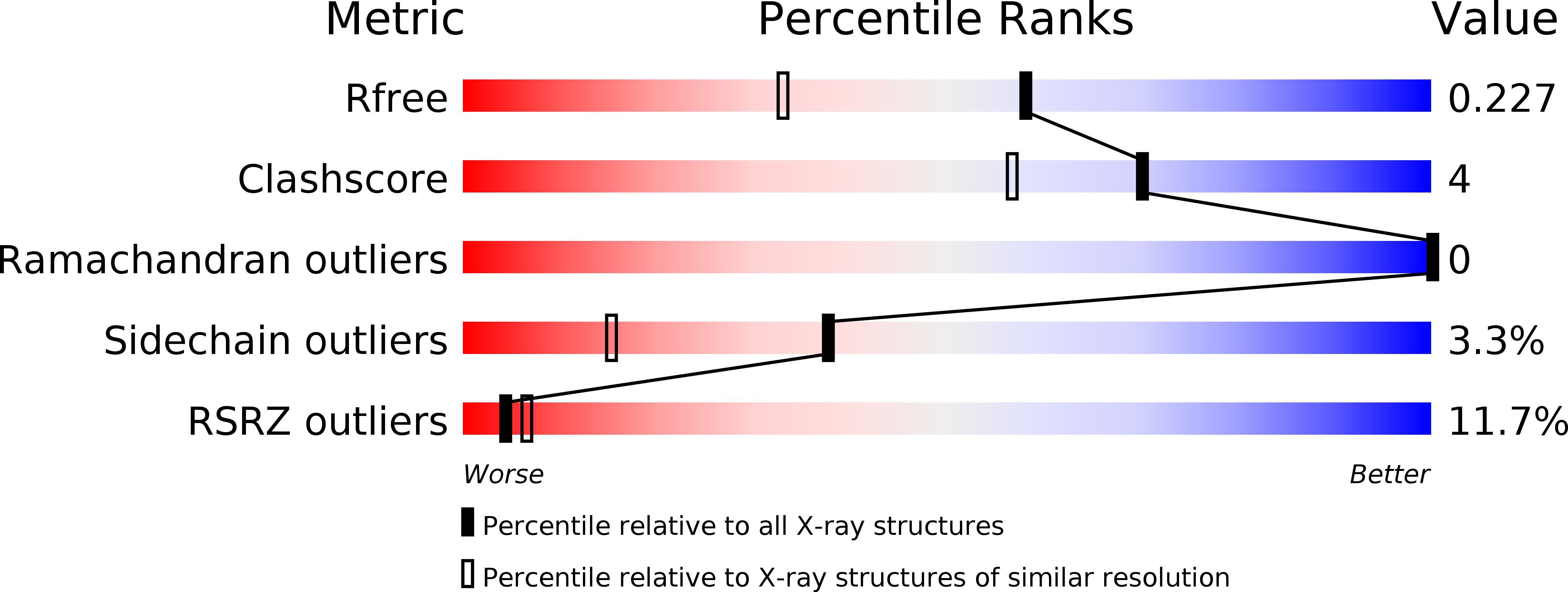
R-Value Free:
0.22
R-Value Work:
0.18
R-Value Observed:
0.18
Space Group:
C 1 2 1