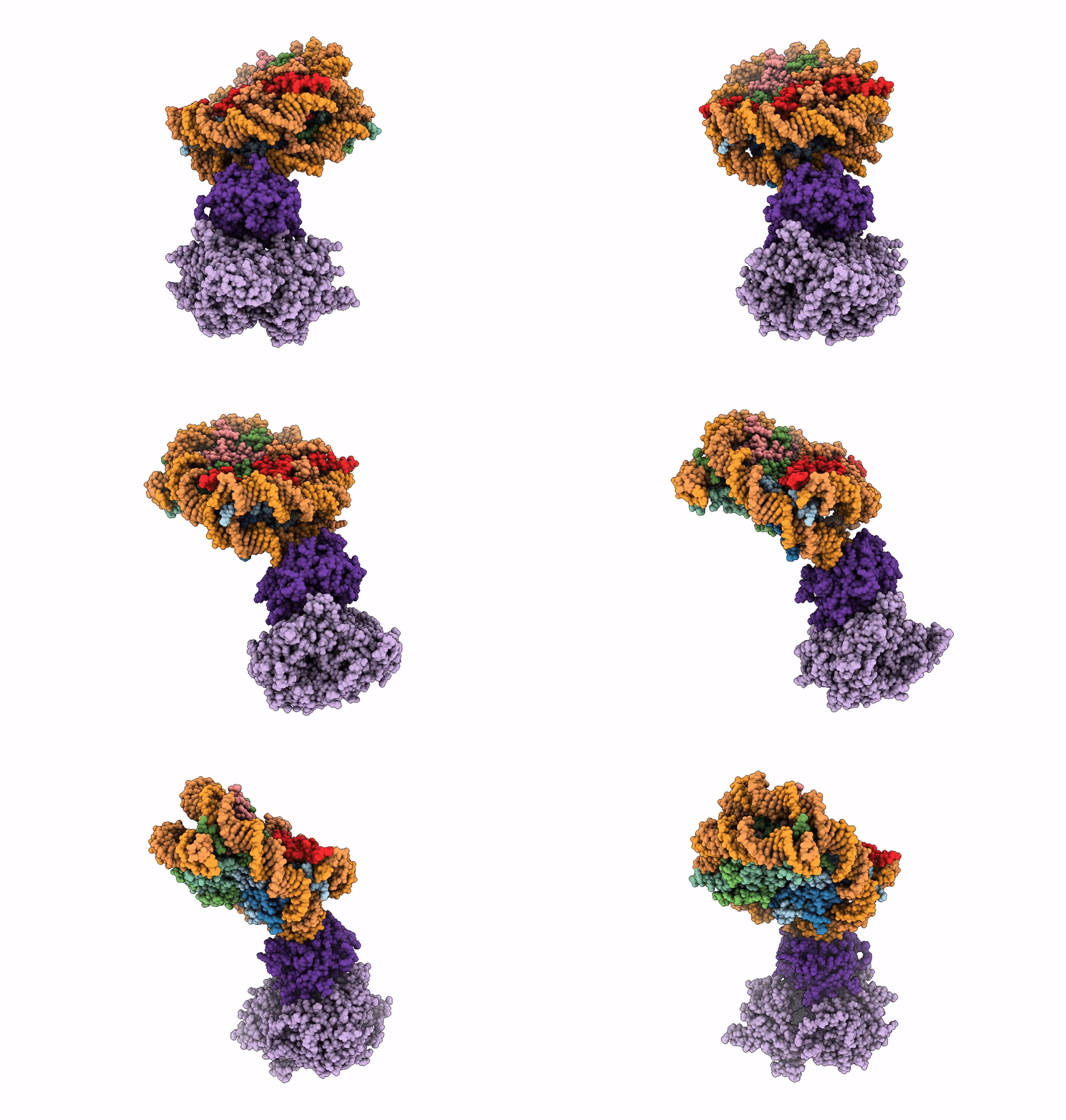
6R92 image
Deposition Date 2019-04-02
Release Date 2019-06-12
Last Version Date 2024-05-22
Entry Detail
PDB ID:
6R92
Title:
Cryo-EM structure of NCP-THF2(+1)-UV-DDB class B
Biological Source:
Source Organism:
Homo sapiens (Taxon ID: 9606)
Host Organism:
Method Details:
Experimental Method:
Resolution:
4.80 Å
Aggregation State:
PARTICLE
Reconstruction Method:
SINGLE PARTICLE
Macromolecular Entities
Polymer Type:polypeptide(L)
Molecule:Histone H3.1
Gene (Uniprot):H3C1, H3C2, H3C3, H3C4, H3C6, H3C7, H3C8, H3C10, H3C11, H3C12
Chain IDs:D (auth: A), H (auth: E)
Chain Length:139
Number of Molecules:2
Biological Source:Homo sapiens
Polymer Type:polypeptide(L)
Molecule:Histone H4
Gene (Uniprot):H4C1, H4C2, H4C3, H4C4, H4C5, H4C6, H4C8, H4C9, H4C11, H4C12, H4C13, H4C14, H4C15, H4C16
Chain IDs:E (auth: B), I (auth: F)
Chain Length:106
Number of Molecules:2
Biological Source:Homo sapiens
Polymer Type:polypeptide(L)
Molecule:Histone H2A type 1-B/E
Gene (Uniprot):H2AC4, H2AC8
Chain IDs:F (auth: C), J (auth: G)
Chain Length:133
Number of Molecules:2
Biological Source:Homo sapiens
Polymer Type:polypeptide(L)
Molecule:Histone H2B type 1-J
Gene (Uniprot):H2BC11
Chain IDs:G (auth: D), K (auth: H)
Chain Length:129
Number of Molecules:2
Biological Source:Homo sapiens
Polymer Type:polydeoxyribonucleotide
Molecule:Human alpha-satellite DNA (145-MER)
Chain IDs:A (auth: I)
Chain Length:145
Number of Molecules:1
Biological Source:Homo sapiens
Polymer Type:polydeoxyribonucleotide
Molecule:Human alpha-satellite DNA (145-MER) with abasic sites at positions 93-94
Chain IDs:B (auth: J)
Chain Length:145
Number of Molecules:1
Biological Source:Homo sapiens
Polymer Type:polypeptide(L)
Molecule:DNA damage-binding protein 1,DNA damage-binding protein 1
Gene (Uniprot):DDB1
Chain IDs:L (auth: K)
Chain Length:1163
Number of Molecules:1
Biological Source:Homo sapiens
Polymer Type:polypeptide(L)
Molecule:DNA damage-binding protein 2
Gene (Uniprot):DDB2
Chain IDs:C (auth: L)
Chain Length:450
Number of Molecules:1
Biological Source:Homo sapiens
Primary Citation
DNA damage detection in nucleosomes involves DNA register shifting.
Nature 571 79 84 (2019)
PMID: 31142837 DOI: 10.1038/s41586-019-1259-3

Abstact

Access to DNA packaged in nucleosomes is critical for gene regulation, DNA replication and DNA repair. In humans, the UV-damaged DNA-binding protein (UV-DDB) complex detects UV-light-induced pyrimidine dimers throughout the genome; however, it remains unknown how these lesions are recognized in chromatin, in which nucleosomes restrict access to DNA. Here we report cryo-electron microscopy structures of UV-DDB bound to nucleosomes bearing a 6-4 pyrimidine-pyrimidone dimer or a DNA-damage mimic in various positions. We find that UV-DDB binds UV-damaged nucleosomes at lesions located in the solvent-facing minor groove without affecting the overall nucleosome architecture. In the case of buried lesions that face the histone core, UV-DDB changes the predominant translational register of the nucleosome and selectively binds the lesion in an accessible, exposed position. Our findings explain how UV-DDB detects occluded lesions in strongly positioned nucleosomes, and identify slide-assisted site exposure as a mechanism by which high-affinity DNA-binding proteins can access otherwise occluded sites in nucleosomal DNA.

Legend

Protein

Chemical

Disease

Primary Citation of related structures