6R63 image
Deposition Date 2019-03-26
Release Date 2019-10-02
Last Version Date 2024-10-09
Entry Detail
PDB ID:
6R63
Keywords:
Title:
Crystal structure of indoleamine 2,3-dioxygenase 1 (IDO1) in complex with ferric heme and MMG-0358
Biological Source:
Source Organism:
Homo sapiens (Taxon ID: 9606)
Method Details:
Experimental Method:
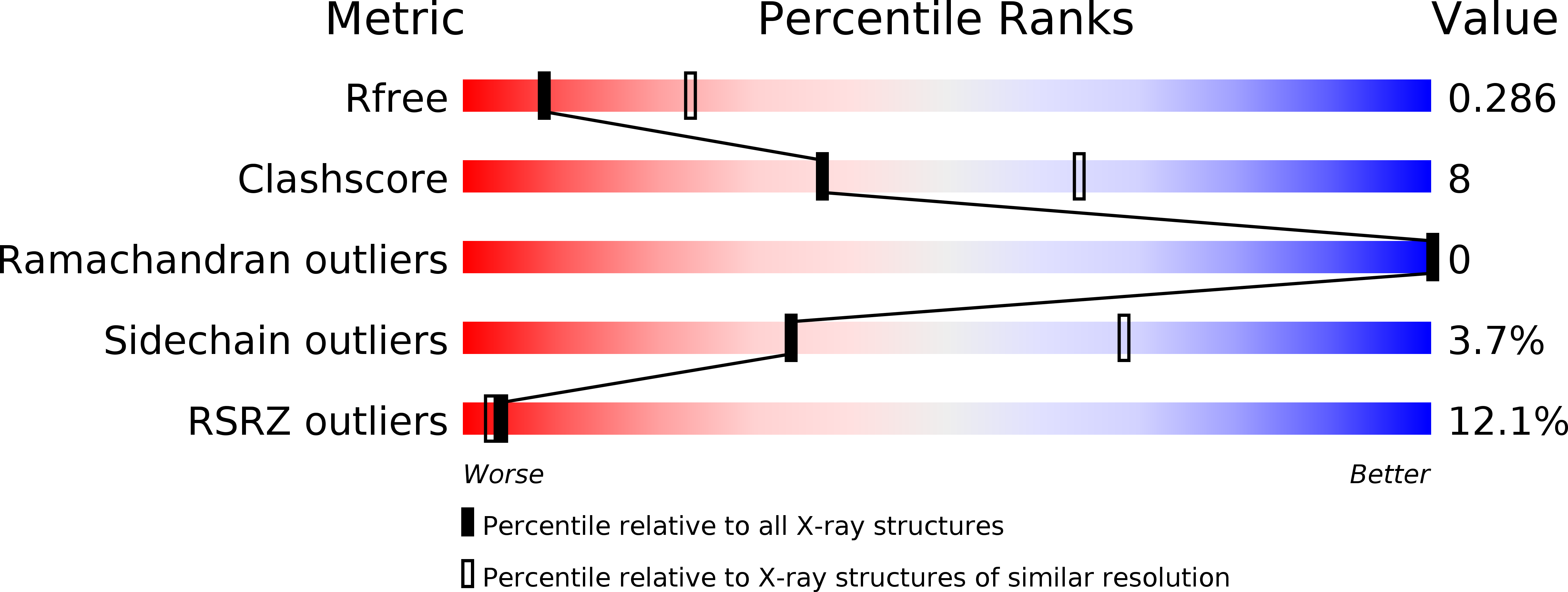
Resolution:
2.89 Å
R-Value Free:
0.28
R-Value Work:
0.23
R-Value Observed:
0.23
Space Group:
P 21 21 21
Macromolecular Entities
Structures with similar UniProt ID
Protein Blast
Polymer Type:polypeptide(L)
Molecule:Indoleamine 2,3-dioxygenase 1
Gene (Uniprot):IDO1
Chain IDs:A, B
Chain Length:423
Number of Molecules:2
Biological Source:Homo sapiens
Primary Citation
Inhibition Mechanisms of Indoleamine 2,3-Dioxygenase 1 (IDO1).
J.Med.Chem. 62 8784 8795 (2019)
PMID: 31525930 DOI: 10.1021/acs.jmedchem.9b00942

Abstact

Indoleamine 2,3-dioxygenase 1 (IDO1) catalyzes the rate-limiting step in the kynurenine pathway of tryptophan metabolism, which is involved in immunity, neuronal function, and aging. Its implication in pathologies such as cancer and neurodegenerative diseases has stimulated the development of IDO1 inhibitors. However, negative phase III clinical trial results of the IDO1 inhibitor epacadostat in cancer immunotherapy call for a better understanding of the role and the mechanisms of IDO1 inhibition. In this work, we investigate the molecular inhibition mechanisms of four known IDO1 inhibitors and of two quinones in detail, using different experimental and computational approaches. We also determine for the first time the X-ray structure of the highly efficient 1,2,3-triazole inhibitor MMG-0358. Based on our results and a comprehensive literature overview, we propose a classification scheme for IDO1 inhibitors according to their inhibition mechanism, which will be useful for further developments in the field.

Legend

Protein

Chemical

Disease

Primary Citation of related structures