6R4P image
Entry Detail
PDB ID:
6R4P
EMDB ID:
Title:
Structure of a soluble domain of adenylyl cyclase bound to an activated stimulatory G protein
Biological Source:
Source Organism:
Host Organism:
PDB Version:
Deposition Date:
2019-03-22
Release Date:
2019-05-08
Method Details:
Experimental Method:
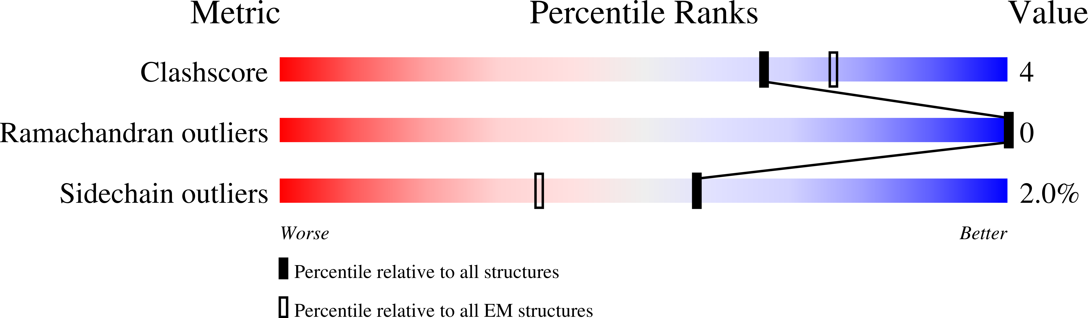
Resolution:
3.10 Å
Aggregation State:
PARTICLE
Reconstruction Method:
SINGLE PARTICLE
Macromolecular Entities
Polymer Type:polypeptide(L)
Description:Adenylate cyclase 9
Chain IDs:A
Chain Length:1637
Number of Molecules:1
Biological Source:Bos taurus
Polymer Type:polypeptide(L)
Description:Guanine nucleotide-binding protein G(s) subunit alpha isoforms short
Chain IDs:B
Chain Length:404
Number of Molecules:1
Biological Source:Bos taurus
Primary Citation
The structure of a membrane adenylyl cyclase bound to an activated stimulatory G protein.
Science 364 389 394 (2019)
PMID: 31023924 DOI: 10.1126/science.aav0778

Abstact

Membrane-integral adenylyl cyclases (ACs) are key enzymes in mammalian heterotrimeric GTP-binding protein (G protein)-dependent signal transduction, which is important in many cellular processes. Signals received by the G protein-coupled receptors are conveyed to ACs through G proteins to modulate the levels of cellular cyclic adenosine monophosphate (cAMP). Here, we describe the cryo-electron microscopy structure of the bovine membrane AC9 bound to an activated G protein αs subunit at 3.4-angstrom resolution. The structure reveals the organization of the membrane domain and helical domain that spans between the membrane and catalytic domains of AC9. The carboxyl-terminal extension of the catalytic domain occludes both the catalytic and the allosteric sites of AC9, inducing a conformation distinct from the substrate- and activator-bound state, suggesting a regulatory role in cAMP production.

Legend

Protein

Chemical

Disease

Primary Citation of related structures