6R2N image
Deposition Date 2019-03-18
Release Date 2019-10-16
Last Version Date 2024-01-24
Entry Detail
PDB ID:
6R2N
Keywords:
Title:
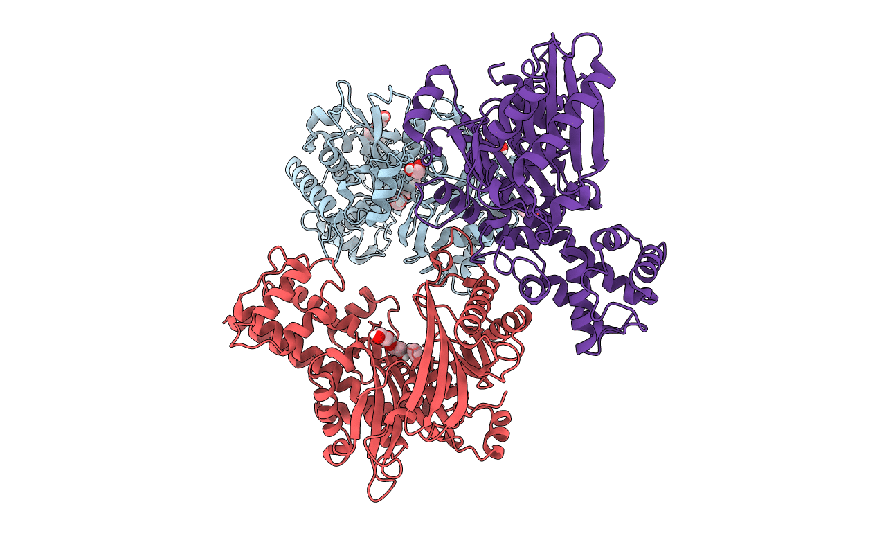
Crystal structure of KlGlk1 glucokinase from Kluyveromyces lactis
Biological Source:
Source Organism:
Method Details:
Experimental Method:
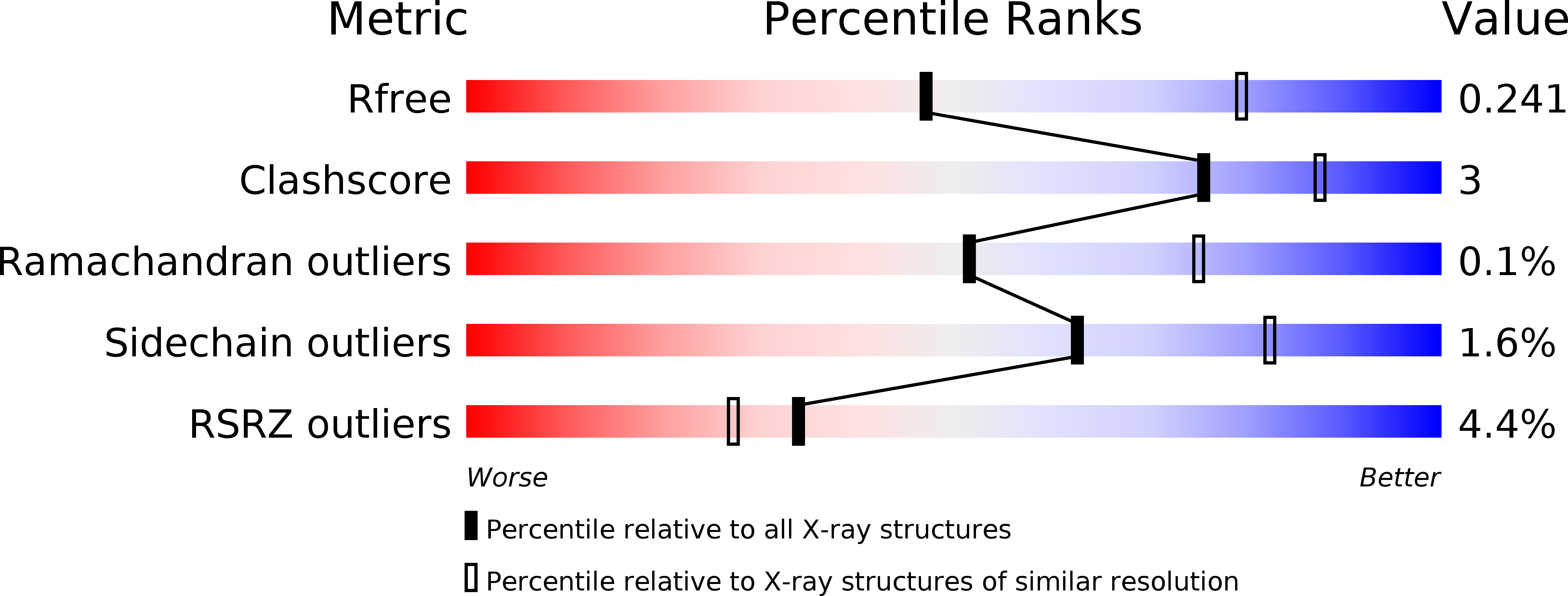
Resolution:
2.60 Å
R-Value Free:
0.24
R-Value Work:
0.20
R-Value Observed:
0.20
Space Group:
C 2 2 21
Macromolecular Entities
Structures with similar UniProt ID
Protein Blast
Polymer Type:polypeptide(L)
Molecule:Glucokinase-1
Gene (Uniprot):GLK1
Chain IDs:A, B, C
Chain Length:475
Number of Molecules:3
Biological Source:Kluyveromyces lactis
Primary Citation
Crystal Structure of Kluyveromyces lactis Glucokinase ( Kl Glk1).
Int J Mol Sci 20 ? ? (2019)
PMID: 31569356 DOI: 10.3390/ijms20194821

Abstact

Glucose phosphorylating enzymes are crucial in the regulation of basic cellular processes, including metabolism and gene expression. Glucokinases and hexokinases provide a pool of phosphorylated glucose in an adenosine diphosphate (ADP)- and ATP-dependent manner to shape the cell metabolism. The glucose processing enzymes from Kluyveromyces lactis are poorly characterized despite the emerging contribution of this yeast strain to industrial and laboratory scale biotechnology. The first reports on K. lactis glucokinase (KlGlk1) positioned the enzyme as an essential component required for glucose signaling. Nevertheless, no biochemical and structural information was available until now. Here, we present the first crystal structure of KlGlk1 together with biochemical characterization, including substrate specificity and enzyme kinetics. Additionally, comparative analysis of the presented structure and the prior structures of lactis hexokinase (KlHxk1) demonstrates the potential transitions between open and closed enzyme conformations upon ligand binding.

Legend

Protein

Chemical

Disease

Primary Citation of related structures