6QXA image
Entry Detail
PDB ID:
6QXA
Title:
Structure of membrane bound pyrophosphatase from Thermotoga maritima in complex with imidodiphosphate and N-[(2-amino-6-benzothiazolyl)methyl]-1H-indole-2-carboxamide (ATC)
Biological Source:
Source Organism:
PDB Version:
Deposition Date:
2019-03-07
Release Date:
2019-04-10
Method Details:
Experimental Method:
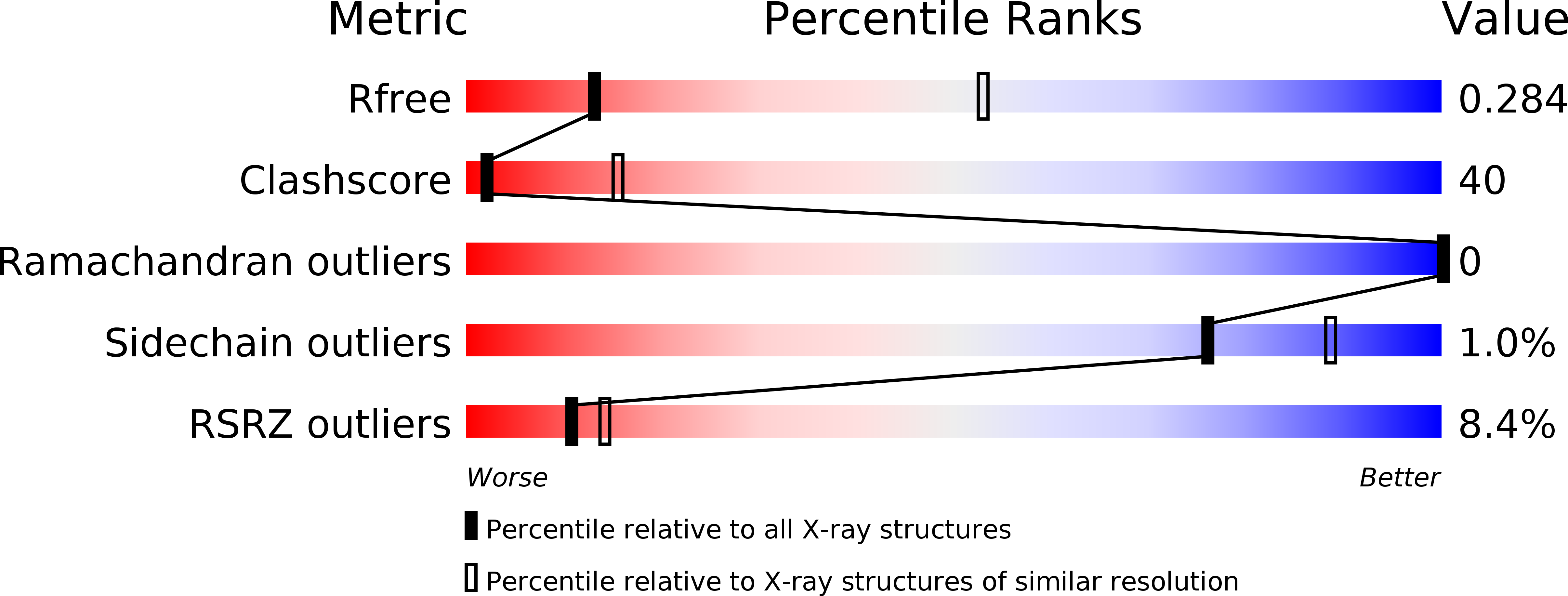
Resolution:
3.41 Å
R-Value Free:
0.28
R-Value Work:
0.22
R-Value Observed:
0.23
Space Group:
P 21 21 21
Macromolecular Entities
Polymer Type:polypeptide(L)
Description:K(+)-stimulated pyrophosphate-energized sodium pump
Chain IDs:A, B, C, D
Chain Length:735
Number of Molecules:4
Biological Source:Thermotoga maritima MSB8
Primary Citation
Asymmetry in catalysis byThermotoga maritimamembrane-bound pyrophosphatase demonstrated by a nonphosphorus allosteric inhibitor.
Sci Adv 5 eaav7574 eaav7574 (2019)
PMID: 31131322 DOI: 10.1126/sciadv.aav7574

Abstact

Membrane-bound pyrophosphatases are homodimeric integral membrane proteins that hydrolyze pyrophosphate into orthophosphates, coupled to the active transport of protons or sodium ions across membranes. They are important in the life cycle of bacteria, archaea, plants, and parasitic protists, but no homologous proteins exist in vertebrates, making them a promising drug target. Here, we report the first nonphosphorus allosteric inhibitor of the thermophilic bacterium Thermotoga maritima membrane-bound pyrophosphatase and its bound structure together with the substrate analog imidodiphosphate. The unit cell contains two protein homodimers, each binding a single inhibitor dimer near the exit channel, creating a hydrophobic clamp that inhibits the movement of β-strand 1-2 during pumping, and thus prevents the hydrophobic gate from opening. This asymmetry of inhibitor binding with respect to each homodimer provides the first clear structural demonstration of asymmetry in the catalytic cycle of membrane-bound pyrophosphatases.

Legend

Protein

Chemical

Disease

Primary Citation of related structures