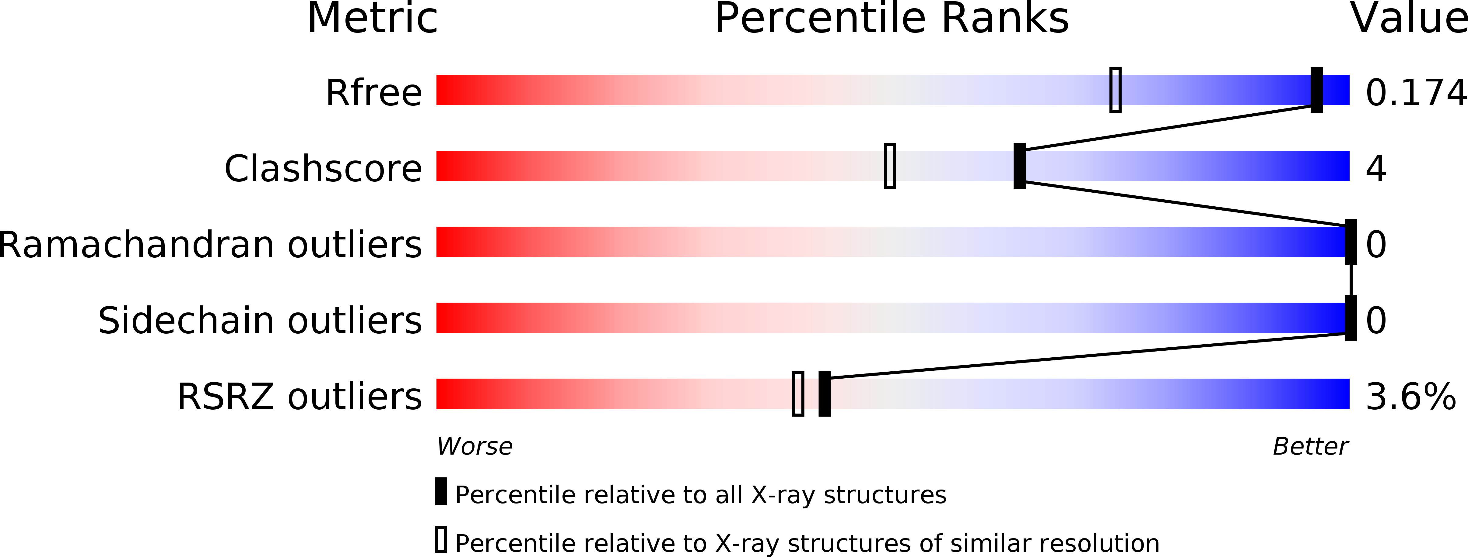
6QW3 image
Deposition Date 2019-03-05
Release Date 2019-08-28
Last Version Date 2024-11-13
Entry Detail
PDB ID:
6QW3
Title:
Calcium-bound gelsolin domain 2
Biological Source:
Source Organism:
Homo sapiens (Taxon ID: 9606)
Host Organism:
Method Details:
Experimental Method:
Resolution:
1.30 Å
R-Value Free:
0.17
R-Value Work:
0.15
R-Value Observed:
0.15
Space Group:
P 2 21 21
Macromolecular Entities
Structures with similar UniProt ID
Protein Blast
Polymer Type:polypeptide(L)
Molecule:Gelsolin
Gene (Uniprot):GSN
Chain IDs:A
Chain Length:119
Number of Molecules:1
Biological Source:Homo sapiens
Ligand Molecules
Primary Citation
High-resolution crystal structure of gelsolin domain 2 in complex with the physiological calcium ion.
Biochem.Biophys.Res.Commun. 518 94 99 (2019)
PMID: 31416615 DOI: 10.1016/j.bbrc.2019.08.013

Abstact

The second domain of gelsolin (G2) hosts mutations responsible for a hereditary form of amyloidosis. The active form of gelsolin is Ca2+-bound; it is also a dynamic protein, hence structural biologists often rely on the study of the isolated G2. However, the wild type G2 structure that have been used so far in comparative studies is bound to a crystallographic Cd2+, in lieu of the physiological calcium. Here, we report the wild type structure of G2 in complex with Ca2+ highlighting subtle ion-dependent differences. Previous findings on different G2 mutations are also briefly revised in light of these results.

Legend

Protein

Chemical

Disease

Primary Citation of related structures