6QVT image
Deposition Date 2019-03-04
Release Date 2020-06-10
Last Version Date 2024-11-20
Entry Detail
PDB ID:
6QVT
Keywords:
Title:
CMP-Sialic acid bound structure of the human wild type Beta-galactoside alpha-2,6-sialyltransferase 1 (ST6Gal1)
Biological Source:
Source Organism:
Homo sapiens (Taxon ID: 9606)
Method Details:
Experimental Method:
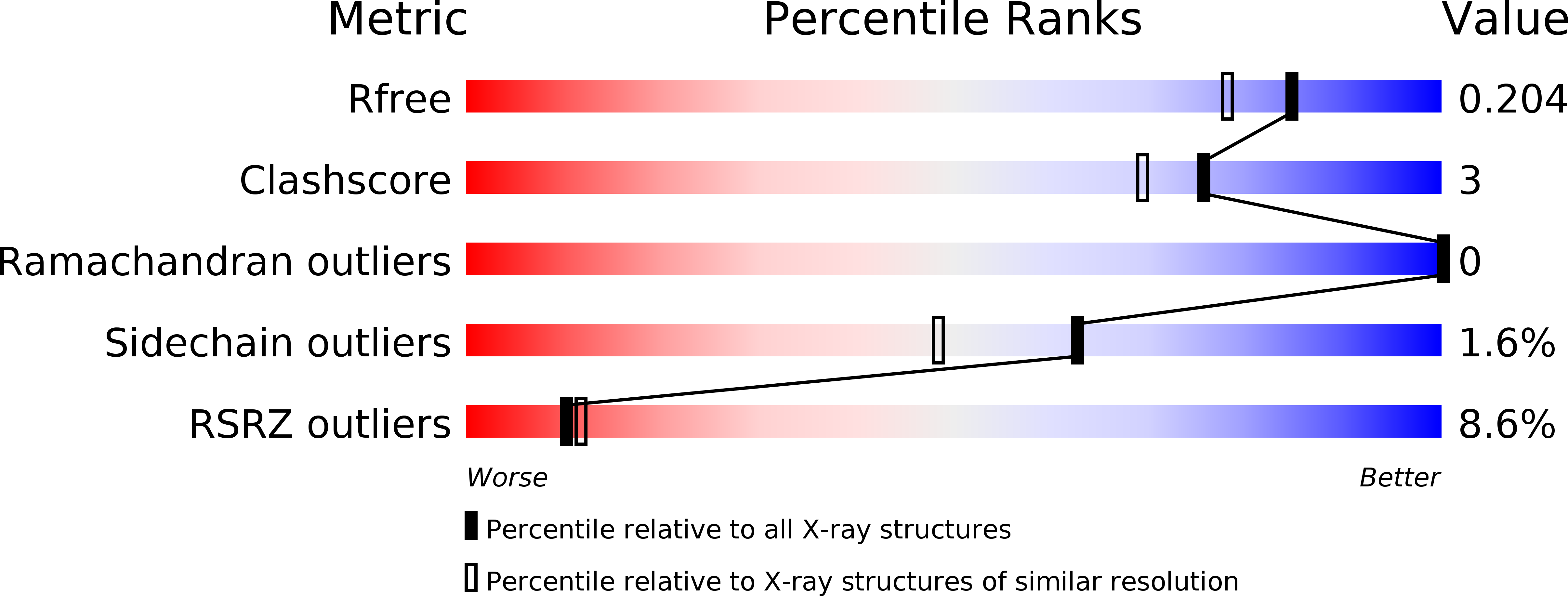
Resolution:
1.70 Å
R-Value Free:
0.20
R-Value Work:
0.18
R-Value Observed:
0.18
Space Group:
C 1 2 1
Macromolecular Entities
Polymer Type:polypeptide(L)
Molecule:Beta-galactoside alpha-2,6-sialyltransferase 1
Gene (Uniprot):ST6GAL1
Chain IDs:A, B
Chain Length:275
Number of Molecules:2
Biological Source:Homo sapiens
Primary Citation
Unliganded and CMP-Neu5Ac bound structures of human alpha-2,6-sialyltransferase ST6Gal I at high resolution.
J.Struct.Biol. 212 107628 107628 (2020)
PMID: 32971290 DOI: 10.1016/j.jsb.2020.107628

Abstact

Sialic acid residues found as terminal monosaccharides in various types of glycan chains in cell surface glycoproteins and glycolipids have been identified as important contributors of cell-cell interactions in normal vs. abnormal cellular behavior and are pivotal in diseases such as cancers. In vertebrates, sialic acids are attached to glycan chains by a conserved subset of sialyltransferases with different enzymatic and substrate specificities. ST6Gal I is a sialyltransferase using activated CMP-sialic acids as donor substrates to catalyze the formation of a α2,6-glycosidic bond between the sialic acid residue and the acceptor disaccharide LacNAc. Understanding sialyltransferases at the molecular and structural level shed light into their function. We present here two human ST6Gal I structures, which show for the first time the enzyme in the unliganded state and with the full donor substrate CMP-Neu5Ac bound. Comparison of these structures reveal flexibility of the catalytic loop, since in the unliganded structure Tyr354 adopts a conformation seen also as an alternate conformation in the substrate bound structure. CMP-Neu5Ac is bound with the side chain at C5 of the sugar residue directed outwards at the surface of the protein. Furthermore, the exact binding mode of the sialic acid moiety of the substrate directly involves sialylmotifs L, S and III and positions the sialylmotif VS in the immediate vicinity. We also present a model for the ternary complex of ST6Gal I with both the donor and the acceptor substrates.

Legend

Protein

Chemical

Disease

Primary Citation of related structures