Deposition Date
2019-02-20
Release Date
2019-05-22
Last Version Date
2024-11-13
Entry Detail
PDB ID:
6QS1
Keywords:
Title:
Crystal structure of human Angiotensin-1 converting enzyme N-domain in complex with BPPb
Biological Source:
Source Organism(s):
Homo sapiens (Taxon ID: 9606)
Gloydius blomhoffii (Taxon ID: 242054)
Gloydius blomhoffii (Taxon ID: 242054)
Expression System(s):
Method Details:
Experimental Method:
Resolution:
1.80 Å
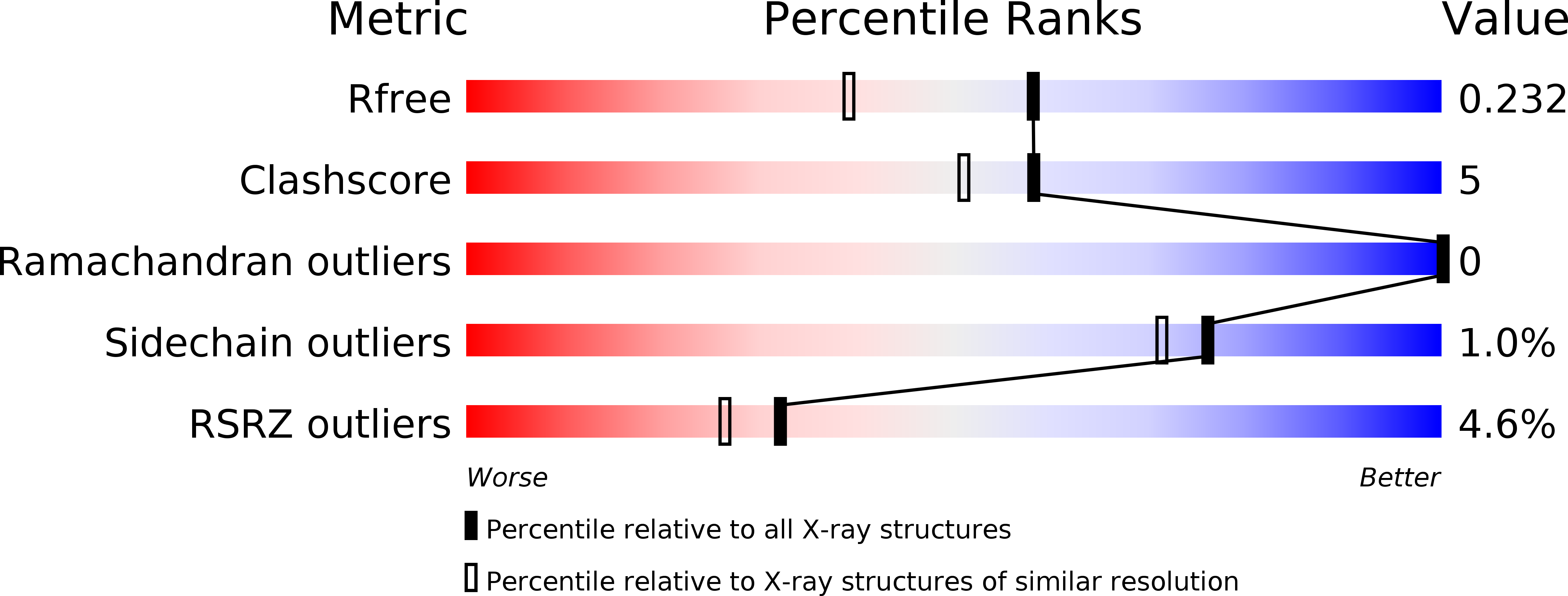
R-Value Free:
0.23
R-Value Work:
0.20
R-Value Observed:
0.20
Space Group:
P 1