Deposition Date
2018-12-28
Release Date
2019-08-21
Last Version Date
2024-11-13
Entry Detail
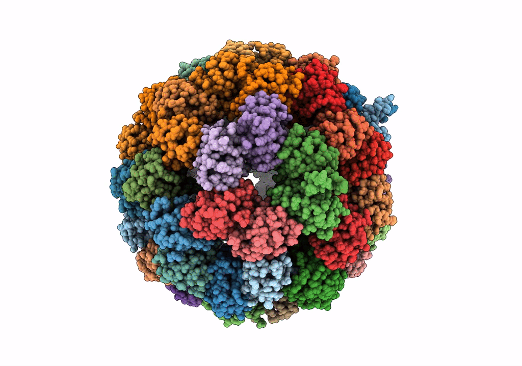
Biological Source:
Source Organism(s):
Listeria monocytogenes EGD-e (Taxon ID: 169963)
Expression System(s):
Method Details:
Experimental Method:
Resolution:
4.21 Å
Aggregation State:
PARTICLE
Reconstruction Method:
SINGLE PARTICLE