6Q5U image
Deposition Date 2018-12-09
Release Date 2019-06-19
Last Version Date 2024-05-15
Entry Detail
PDB ID:
6Q5U
Keywords:
Title:
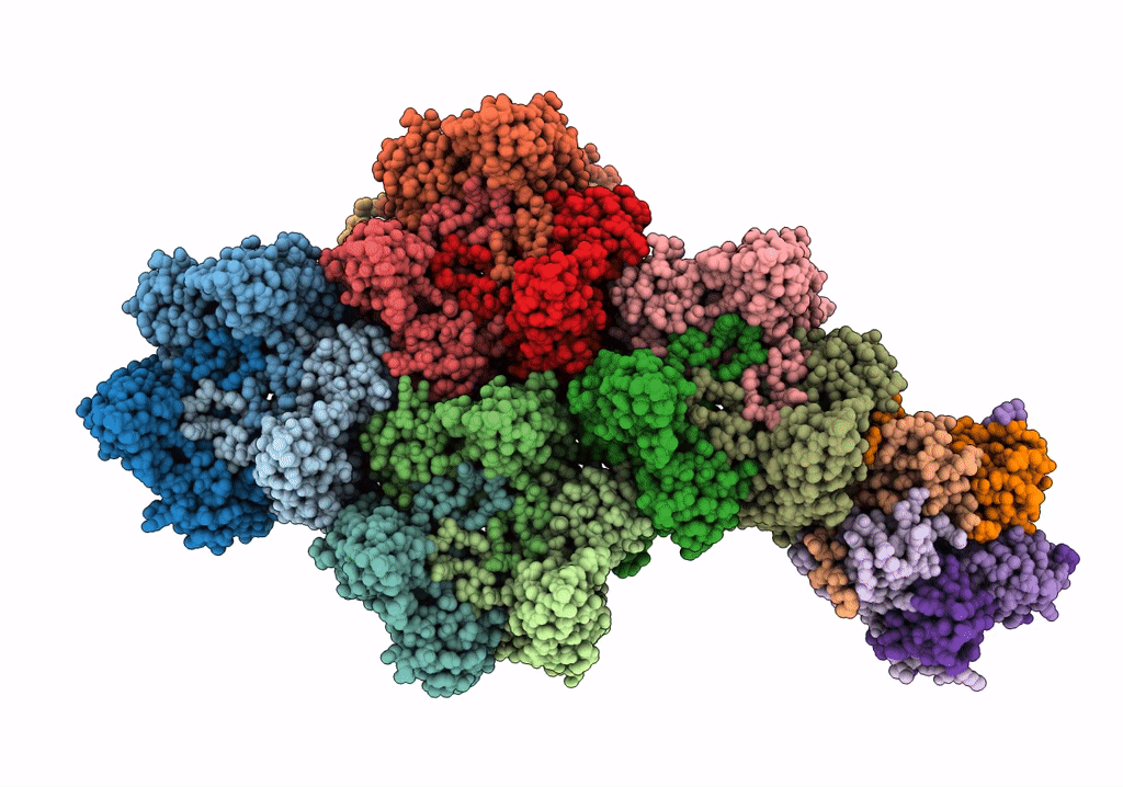
High resolution electron cryo-microscopy structure of the bacteriophage PR772
Biological Source:
Source Organism:
Method Details:
Experimental Method:
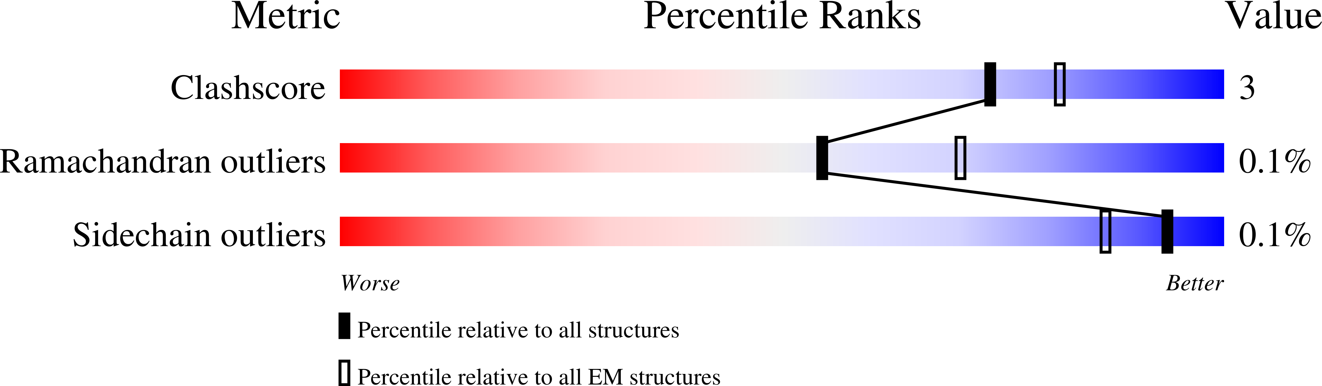
Resolution:
2.75 Å
Aggregation State:
PARTICLE
Reconstruction Method:
SINGLE PARTICLE
Macromolecular Entities
Structures with similar UniProt ID
Protein Blast
Polymer Type:polypeptide(L)
Molecule:Major Capsid Protein (P3)
Gene (Uniprot):III
Chain IDs:A, B, C, D, E, F, G, H, I, J, K, L
Chain Length:395
Number of Molecules:12
Biological Source:Enterobacteria phage PR772
Structures with similar UniProt ID
Protein Blast
Polymer Type:polypeptide(L)
Molecule:Minor Capsid Protein (P30)
Gene (Uniprot):XXX
Chain IDs:M
Chain Length:84
Number of Molecules:1
Biological Source:Enterobacteria phage PR772
Structures with similar UniProt ID
Protein Blast
Polymer Type:polypeptide(L)
Molecule:Infectivity Protein (P16)
Gene (Uniprot):XVI
Chain IDs:N
Chain Length:117
Number of Molecules:1
Biological Source:Enterobacteria phage PR772
Structures with similar UniProt ID
Protein Blast
Polymer Type:polypeptide(L)
Molecule:Spike protein
Gene (Uniprot):V
Chain IDs:O, P, Q
Chain Length:340
Number of Molecules:3
Biological Source:Enterobacteria phage PR772
Structures with similar UniProt ID
Protein Blast
Polymer Type:polypeptide(L)
Molecule:Penton protein
Gene (Uniprot):XXXI
Chain IDs:R, S
Chain Length:126
Number of Molecules:2
Biological Source:Enterobacteria phage PR772
Ligand Molecules
Primary Citation
Electron cryo-microscopy of Bacteriophage PR772 reveals the elusive vertex complex and the capsid architecture.
Elife 8 ? ? (2019)
PMID: 31513011 DOI: 10.7554/eLife.48496

Abstact

Bacteriophage PR772, a member of the Tectiviridae family, has a 70 nm diameter icosahedral protein capsid that encapsulates a lipid membrane, dsDNA, and various internal proteins. An icosahedrally averaged CryoEM reconstruction of the wild-type virion and a localized reconstruction of the vertex region reveal the composition and the structure of the vertex complex along with new protein conformations that play a vital role in maintaining the capsid architecture of the virion. The overall resolution of the virion is 2.75 Å, while the resolution of the protein capsid is 2.3 Å. The conventional penta-symmetron formed by the capsomeres is replaced by a large vertex complex in the pseudo T = 25 capsid. All the vertices contain the host-recognition protein, P5; two of these vertices show the presence of the receptor-binding protein, P2. The 3D structure of the vertex complex shows interactions with the viral membrane, indicating a possible mechanism for viral infection.

Legend

Protein

Chemical

Disease

Primary Citation of related structures
Feedback Form
Name
Email
Institute
Feedback