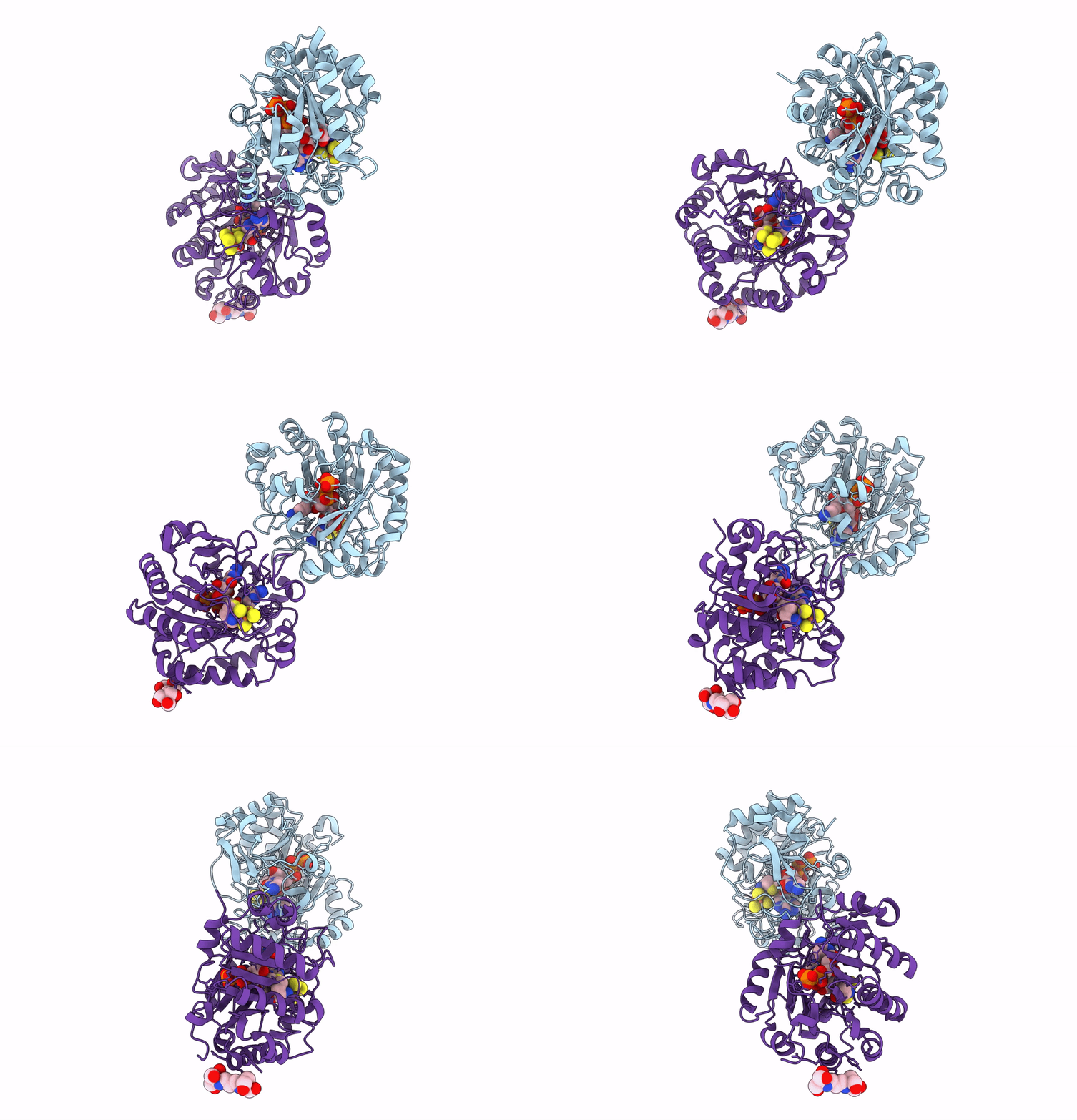
6Q2P image
Deposition Date 2019-08-08
Release Date 2020-01-22
Last Version Date 2023-10-11
Entry Detail
PDB ID:
6Q2P
Keywords:
Title:
Crystal structure of mouse viperin bound to cytidine triphosphate and S-adenosylhomocysteine
Biological Source:
Source Organism:
Mus musculus (Taxon ID: 10090)
Method Details:
Experimental Method:
Resolution:
1.45 Å
R-Value Free:
0.15
R-Value Work:
0.14
R-Value Observed:
0.14
Space Group:
P 21 21 21
Macromolecular Entities
Polymer Type:polypeptide(L)
Molecule:Radical S-adenosyl methionine domain-containing protein 2
Gene (Uniprot):Rsad2
Mutations:E261A, E266A
Chain IDs:A, B
Chain Length:318
Number of Molecules:2
Biological Source:Mus musculus
Primary Citation
Structural Basis of the Substrate Selectivity of Viperin.
Biochemistry 59 652 662 (2020)
PMID: 31917549 DOI: 10.1021/acs.biochem.9b00741

Abstact

Viperin is a radical S-adenosylmethionine (SAM) enzyme that inhibits viral replication by converting cytidine triphosphate (CTP) into 3'-deoxy-3',4'-didehydro-CTP and by additional undefined mechanisms operating through its N- and C-terminal domains. Here, we describe crystal structures of viperin bound to a SAM analogue and CTP or uridine triphosphate (UTP) and report kinetic parameters for viperin-catalyzed reactions with CTP or UTP as substrates. Viperin orients the C4' hydrogen atom of CTP and UTP similarly for abstraction by a 5'-deoxyadenosyl radical, but the uracil moiety introduces unfavorable interactions that prevent tight binding of UTP. Consistently, kcat is similar for CTP and UTP whereas the Km for UTP is much greater. The structures also show that nucleotide binding results in ordering of the C-terminal tail and reveal that this region contains a P-loop that binds the γ-phosphate of the bound nucleotide. Collectively, the results explain the selectivity for CTP and reveal a structural role for the C-terminal tail in binding CTP and UTP.

Legend

Protein

Chemical

Disease

Primary Citation of related structures