6Q2M image
Deposition Date 2019-08-08
Release Date 2019-10-16
Last Version Date 2023-10-11
Entry Detail
PDB ID:
6Q2M
Keywords:
Title:
Crystal structure of Photinus pyralis Luciferase Pps6 mutant in complex with DLSA
Biological Source:
Source Organism:
Method Details:
Experimental Method:
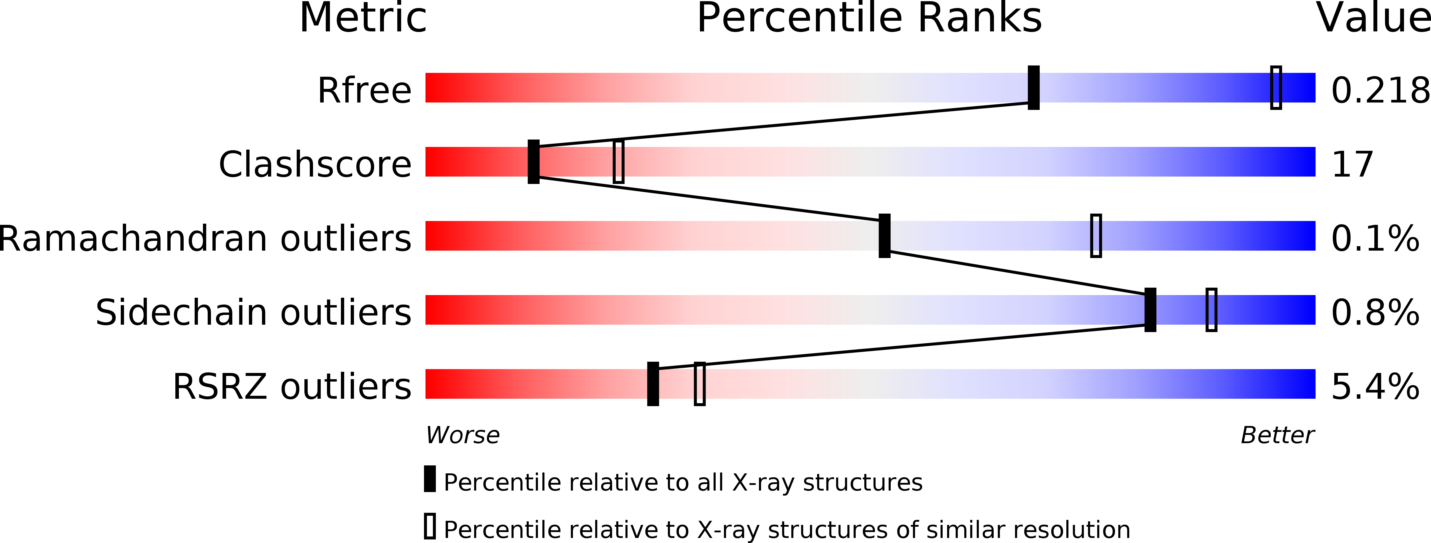
Resolution:
2.75 Å
R-Value Free:
0.21
R-Value Work:
0.19
R-Value Observed:
0.19
Space Group:
P 1 21 1
Macromolecular Entities
Polymer Type:polypeptide(L)
Molecule:Luciferin 4-monooxygenase
Mutations:T214N, A222C, Y255F, S276T, H332N, E354N
Chain IDs:A, B, C
Chain Length:555
Number of Molecules:3
Biological Source:Photinus pyralis
Primary Citation
Mutagenesis and Structural Studies Reveal the Basis for the Activity and Stability Properties That Distinguish thePhotinusLuciferasesscintillansandpyralis.
Biochemistry 58 4293 4303 (2019)
PMID: 31560532 DOI: 10.1021/acs.biochem.9b00719

Abstact

The dazzling yellow-green light emission of the common North American firefly Photinus pyralis and other bioluminescent organisms has provided a wide variety of prominent research applications like reporter gene assays and in vivo imaging methods. While the P. pyralis enzyme has been extensively studied, only recently has a second Photinus luciferase been cloned from the species scintillans. Even though the enzymes share very high sequence identity (89.8%), the color of the light they emit, their specific activity and their stability to heat, pH, and chemical denaturation are quite different with the scintillans luciferase being generally more resistant. Through the construction and evaluation of the properties of chimeric domain swapped, single point, and various combined variants, we have determined that only six amino acid changes are necessary to confer all of the properties of the scintillans enzyme to wild-type P. pyralis luciferase. Altered stability properties were attributed to four of the amino acid changes (T214N/S276T/H332N/E354N), and single mutations each predominantly changed emission color (Y255F) and specific activity (A222C). Results of a crystallographic study of the P. pyralis enzyme containing the six changes (Pps6) provide some insight into the structural basis for some of the documented property differences.

Legend

Protein

Chemical

Disease

Primary Citation of related structures