Deposition Date
2019-07-25
Release Date
2020-06-24
Last Version Date
2024-10-09
Entry Detail
PDB ID:
6PXE
Keywords:
Title:
Crystal structure of the complex between periplasmic domains of antiholin RI and holin T from T4 phage, in P21
Biological Source:
Source Organism(s):
Enterobacteria phage T4 (Taxon ID: 10665)
Escherichia phage vB_EcoM_NBG2 (Taxon ID: 2184699)
Escherichia phage vB_EcoM_NBG2 (Taxon ID: 2184699)
Expression System(s):
Method Details:
Experimental Method:
Resolution:
2.30 Å
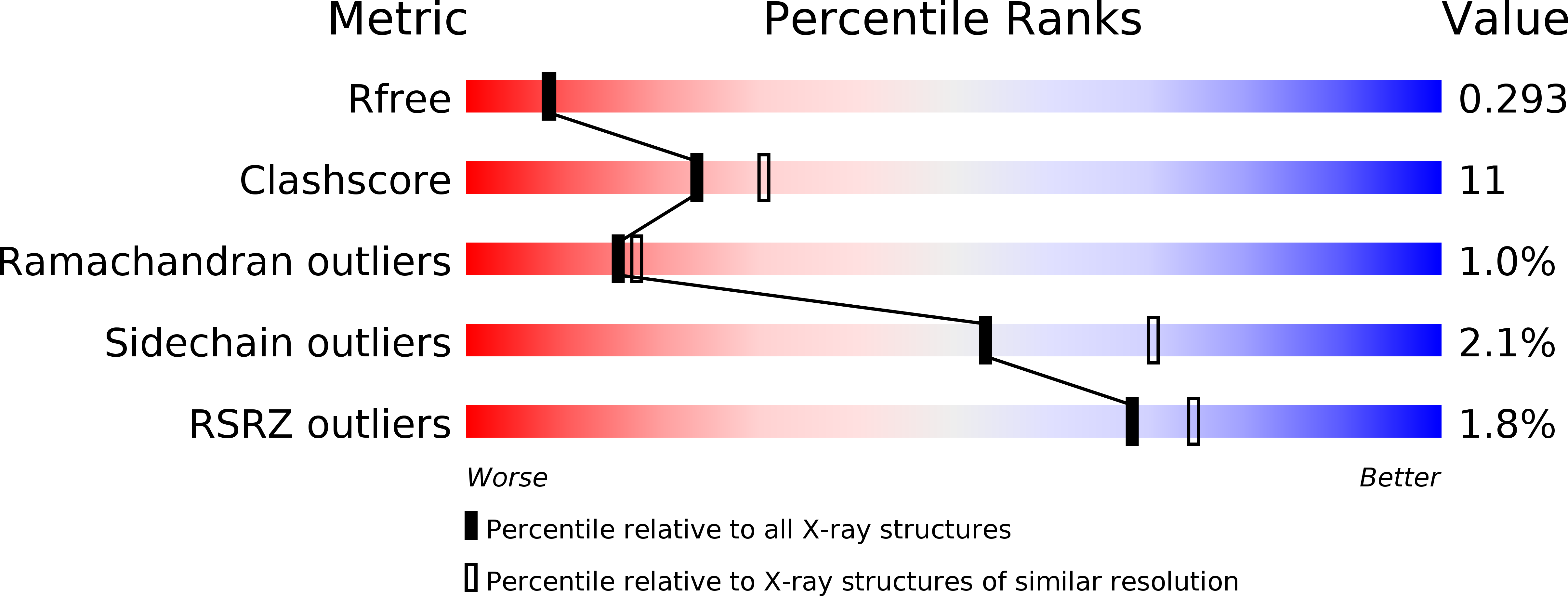
R-Value Free:
0.29
R-Value Work:
0.23
R-Value Observed:
0.23
Space Group:
P 1 21 1