6PW7 image
Deposition Date 2019-07-22
Release Date 2019-11-13
Last Version Date 2024-11-13
Entry Detail
PDB ID:
6PW7
Title:
X-ray crystal structure of C. elegans STIM EF-SAM domain
Biological Source:
Source Organism(s):
Expression System(s):
Method Details:
Experimental Method:
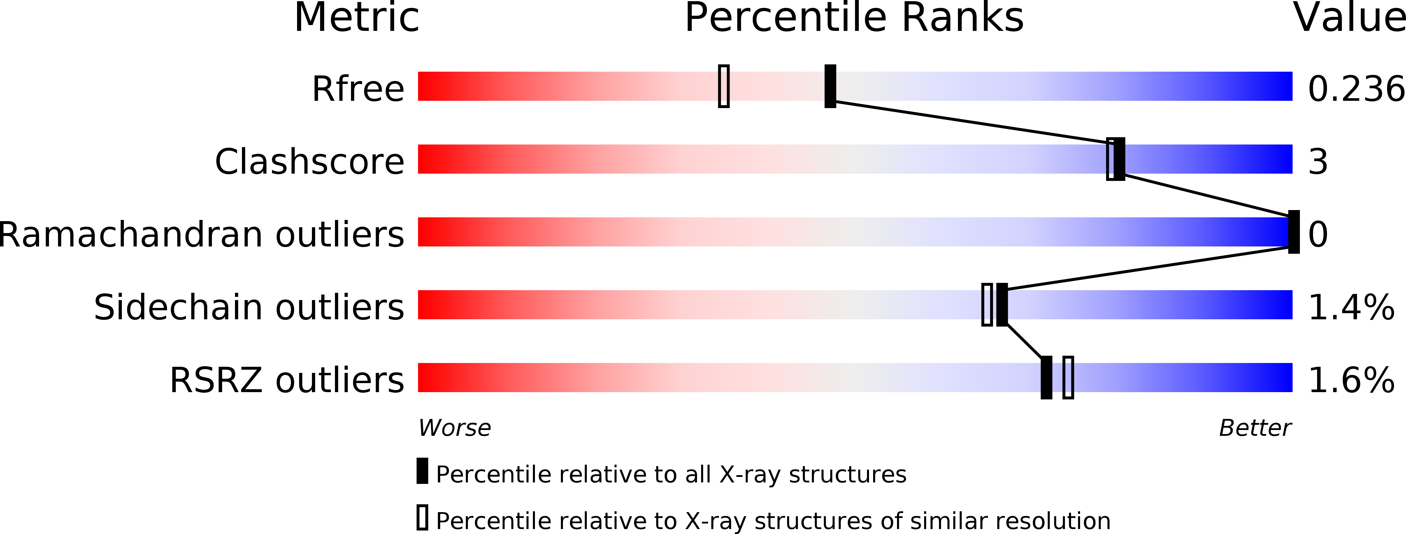
Resolution:
1.89 Å
R-Value Free:
0.23
R-Value Work:
0.18
R-Value Observed:
0.18
Space Group:
C 2 2 2
Macromolecular Entities
Structures with similar UniProt ID
Protein Blast
Polymer Type:polypeptide(L)
Molecule:Stromal interaction molecule 1
Gene (Uniprot):stim-1
Chain IDs:A, B
Chain Length:161
Number of Molecules:2
Biological Source:Caenorhabditis elegans
Modified Residue
Compound ID Chain ID Parent Comp ID Details 2D Image
MSE A MET modified residue
Ligand Molecules
Primary Citation
Coordination of a Single Calcium Ion in the EF-hand Maintains the Off State of the Stromal Interaction Molecule Luminal Domain.
J.Mol.Biol. 432 367 383 (2020)
PMID: 31626806 DOI: 10.1016/j.jmb.2019.10.003

Abstact

Store operated calcium (Ca2+) entry (SOCE) is the process whereby endoplasmic reticulum (ER) Ca2+ store depletion causes Orai1-composed Ca2+ channels on the plasma membrane (PM) to open, mediating a rise in cytosolic Ca2+ levels. Stromal interaction molecules (STIMs) are the proteins that directly sense ER Ca2+ content and gate Orai1 channels due to store depletion. The trigger for STIM activation is Ca2+ unbinding from the ER lumen-oriented domains, which consist of a nonconserved amino (N) terminal region and EF-hand and sterile α motif (SAM) domains (EF-SAM), highly conserved from humans to Caenorhabditis elegans. Solution NMR structures of the human EF-SAM domains have been determined at high Ca2+ concentrations; however, no direct structural view of the Ca2+ binding mode has been elucidated. Further, no atomic resolution data currently exists on EF-SAM at low Ca2+ levels. Here, we determined the X-ray crystal structure of the C. elegans STIM luminal domain, revealing that EF-SAM binds a single Ca2+ ion with pentagonal bipyramidal geometry and an ancillary α-helix formed by the N-terminal region acts as a brace to stabilize EF-SAM. Using solution NMR, we observed EF-hand domain unfolding and a conformational exchange between folded and unfolded states involving the ancillary α-helix and the canonical EF-hand in low Ca2+. Remarkably, we also detected an α-helix (+Ca2+) to β-strand (-Ca2+) transition at the terminal SAM domain α-helix. Collectively, our analyses indicate that one canonically bound Ca2+ ion is sufficient to stabilize the quiescent luminal domain structure, precluding unfolding, conformational exchange, and secondary structure transformation.

Legend

Protein

Chemical

Disease

Primary Citation of related structures
Feedback Form
Name
Email
Institute
Feedback