Deposition Date
2019-07-17
Release Date
2020-07-22
Last Version Date
2024-04-10
Entry Detail
PDB ID:
6PU6
Keywords:
Title:
CobT from Methanocaldococcus jannaschii in complex with Alpha-Ribozole 5'-Phosphate, Nicotinic Acid, and Nicotinic Acid Mononucleotide
Biological Source:
Source Organism(s):
Expression System(s):
Method Details:
Experimental Method:
Resolution:
2.29 Å
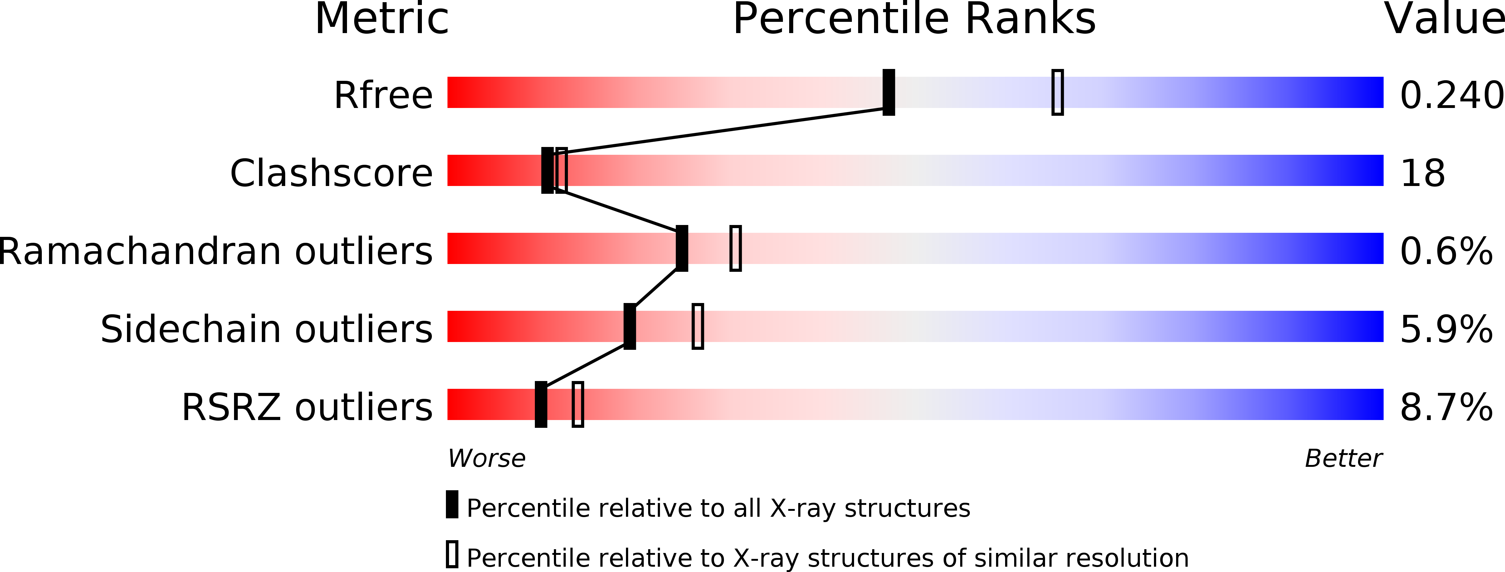
R-Value Free:
0.24
R-Value Work:
0.19
R-Value Observed:
0.19
Space Group:
P 1 21 1