
Deposition Date
2019-07-16
Release Date
2020-05-13
Last Version Date
2024-05-15
Entry Detail
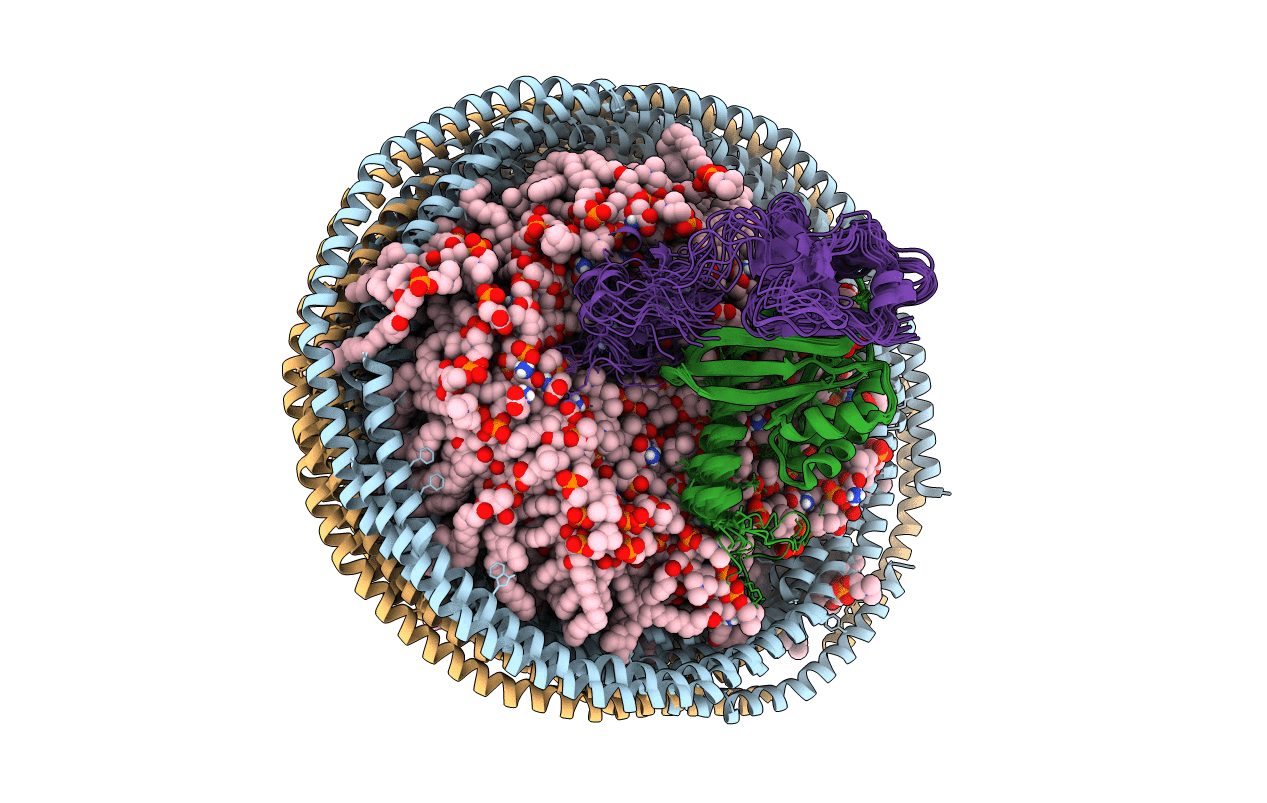
PDB ID:
6PTS
Keywords:
Title:
NMR data-driven model of KRas-GMPPNP:RBD-CRD complex tethered to a nanodisc (state A)
Biological Source:
Source Organism(s):
Homo sapiens (Taxon ID: 9606)
Expression System(s):
Method Details:
Experimental Method:
Conformers Calculated:
3000
Conformers Submitted:
10
Selection Criteria:
structures with the lowest energy
