6PQB image
Deposition Date 2019-07-08
Release Date 2019-10-16
Last Version Date 2023-10-11
Entry Detail
PDB ID:
6PQB
Keywords:
Title:
Crystal structure of aminoglycoside-resistance methyltransferase RmtC bound to S-adenosylhomocysteine (SAH)
Biological Source:
Source Organism:
Method Details:
Experimental Method:
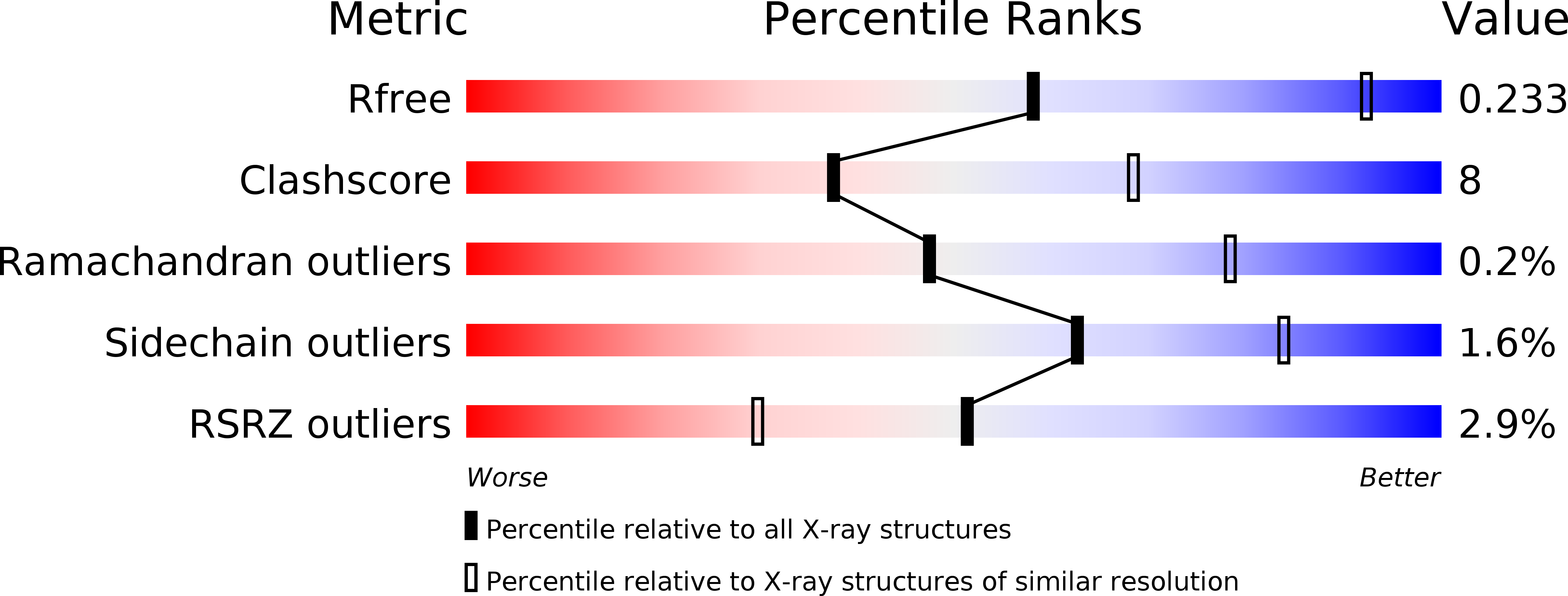
Resolution:
3.14 Å
R-Value Free:
0.23
R-Value Work:
0.20
R-Value Observed:
0.20
Space Group:
P 61
Macromolecular Entities
Structures with similar UniProt ID
Protein Blast
Polymer Type:polypeptide(L)
Molecule:16S rRNA (guanine(1405)-N(7))-methyltransferase
Gene (Uniprot):rmtC
Chain IDs:A, B, C, D
Chain Length:283
Number of Molecules:4
Biological Source:Proteus mirabilis
Ligand Molecules
Primary Citation
Functionally critical residues in the aminoglycoside resistance-associated methyltransferase RmtC play distinct roles in 30S substrate recognition.
J.Biol.Chem. 294 17642 17653 (2019)
PMID: 31594862 DOI: 10.1074/jbc.RA119.011181

Abstact

Methylation of the small ribosome subunit rRNA in the ribosomal decoding center results in exceptionally high-level aminoglycoside resistance in bacteria. Enzymes that methylate 16S rRNA on N7 of nucleotide G1405 (m7G1405) have been identified in both aminoglycoside-producing and clinically drug-resistant pathogenic bacteria. Using a fluorescence polarization 30S-binding assay and a new crystal structure of the methyltransferase RmtC at 3.14 Å resolution, here we report a structure-guided functional study of 30S substrate recognition by the aminoglycoside resistance-associated 16S rRNA (m7G1405) methyltransferases. We found that the binding site for these enzymes in the 30S subunit directly overlaps with that of a second family of aminoglycoside resistance-associated 16S rRNA (m1A1408) methyltransferases, suggesting that both groups of enzymes may exploit the same conserved rRNA tertiary surface for docking to the 30S. Within RmtC, we defined an N-terminal domain surface, comprising basic residues from both the N1 and N2 subdomains, that directly contributes to 30S-binding affinity. In contrast, additional residues lining a contiguous adjacent surface on the C-terminal domain were critical for 16S rRNA modification but did not directly contribute to the binding affinity. The results from our experiments define the critical features of m7G1405 methyltransferase-substrate recognition and distinguish at least two distinct, functionally critical contributions of the tested enzyme residues: 30S-binding affinity and stabilizing a binding-induced 16S rRNA conformation necessary for G1405 modification. Our study sets the scene for future high-resolution structural studies of the 30S-methyltransferase complex and for potential exploitation of unique aspects of substrate recognition in future therapeutic strategies.

Legend

Protein

Chemical

Disease

Primary Citation of related structures