6PON image
Entry Detail
PDB ID:
6PON
Keywords:
Title:
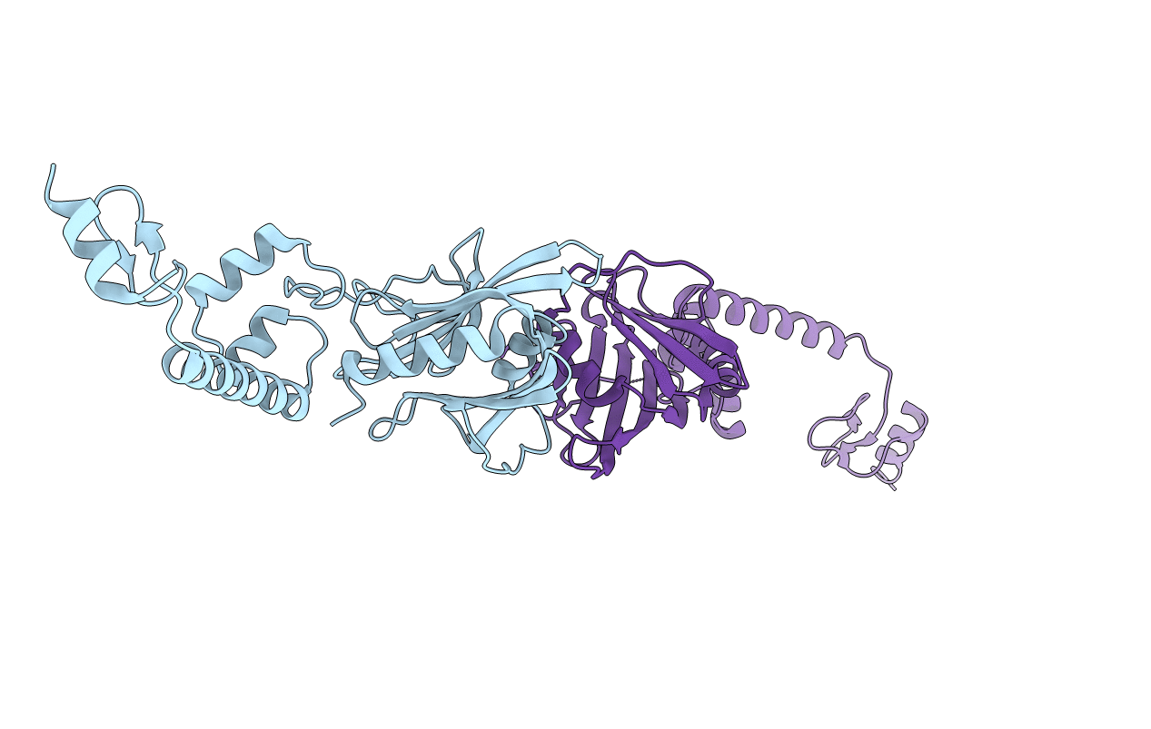
CRYSTAL STRUCTURE OF THE N-TERMINAL DOMAIN OF FIBRONECTIN- BINDING PROTEIN PAVA FROM STREPTOCOCCUS PNEUMONIAE
Biological Source:
Source Organism:
Host Organism:
PDB Version:
Deposition Date:
2019-07-04
Release Date:
2019-07-31
Method Details:
Experimental Method:
Resolution:
2.40 Å
R-Value Free:
0.23
R-Value Work:
0.16
R-Value Observed:
0.17
Space Group:
P 1 21 1
Macromolecular Entities
Polymer Type:polypeptide(L)
Description:Adherence and virulence protein A
Chain IDs:A, B
Chain Length:274
Number of Molecules:2
Biological Source:Streptococcus pneumoniae
Primary Citation
Crystal structure of the N-terminal domain of the fibronectin-binding protein PavA from Streptococcus pneumoniae.
Acta Crystallogr.,Sect.F 75 657 662 (2019)
PMID: 31584015 DOI: 10.1107/S2053230X19012160

Abstact

The Gram-positive bacterium Streptococcus pneumoniae, a major human pathogen, is a regular colonizer of the upper and lower respiratory tracts. Pneumococcal adherence and virulence factor A (PavA), a fibronectin-binding bacterial protein, from S. pneumoniae is an important facilitator of its colonization of host cells. In this study, the crystal structure of the N-terminal domain of PavA (SpPavA-N) determined at a resolution of 2.39 Å is reported. Each monomer of the dimeric protein consists of two domains (domains I and II) and a short α-helix (α6) at the C-terminus that are connected by elongated loops. Comparison of the SpPavA-N structure with that of its homolog from Streptococcus suis (FBPS-N) revealed differences in α5, α6 and the domain II/α6 inter-loop region within domain II. The α5 helix of FBPS-N folds back toward domain I, whereas in SpPavA-N it adopts an elongated rod shape.

Legend

Protein

Chemical

Disease

Primary Citation of related structures