6PL5 image
Entry Detail
PDB ID:
6PL5
Title:
Structural coordination of polymerization and crosslinking by a peptidoglycan synthase complex
Biological Source:
Source Organism:
PDB Version:
Deposition Date:
2019-06-30
Release Date:
2020-03-18
Method Details:
Experimental Method:
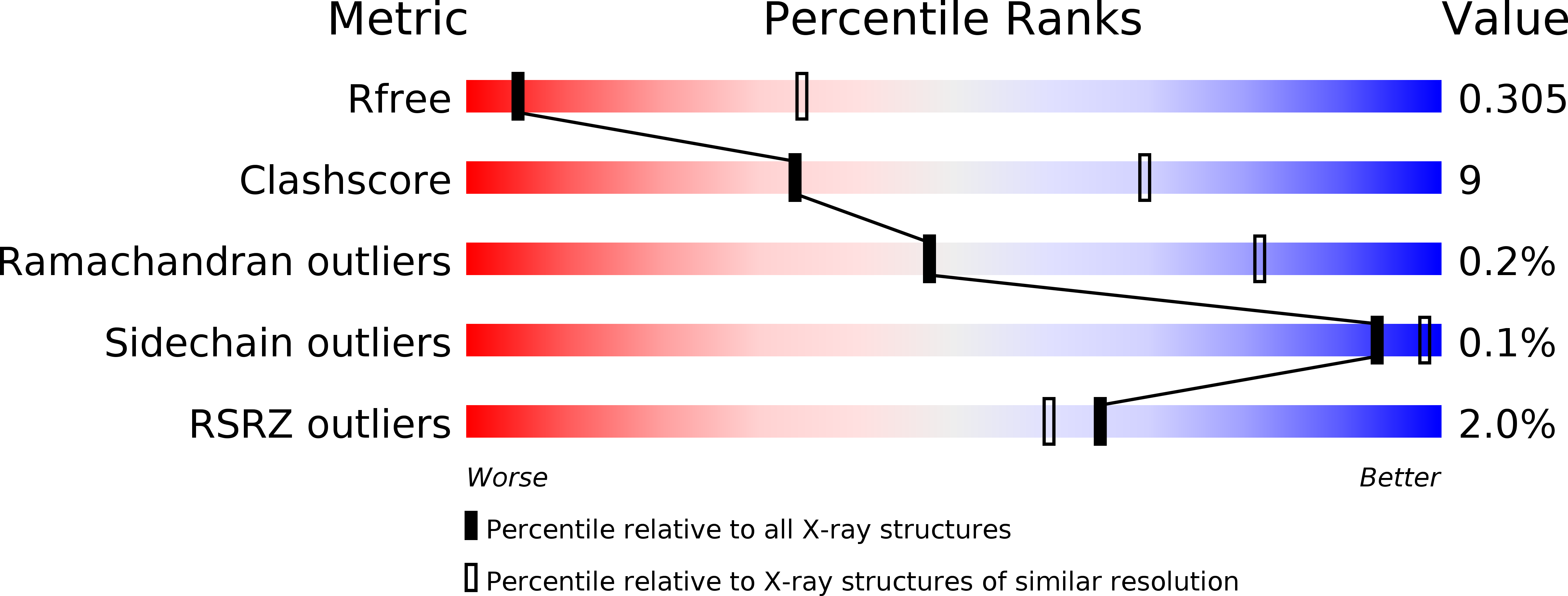
Resolution:
3.50 Å
R-Value Free:
0.30
R-Value Work:
0.28
R-Value Observed:
0.28
Space Group:
P 32 2 1
Macromolecular Entities
Polymer Type:polypeptide(L)
Description:Peptidoglycan glycosyltransferase RodA
Chain IDs:A
Chain Length:377
Number of Molecules:1
Biological Source:Thermus thermophilus HB8
Polymer Type:polypeptide(L)
Description:Penicillin-binding protein 2/cell division protein FtsI
Chain IDs:B
Chain Length:598
Number of Molecules:1
Biological Source:Thermus thermophilus HB8
Polymer Type:polypeptide(L)
Description:Unknown peptide
Chain IDs:C (auth: D)
Chain Length:11
Number of Molecules:1
Biological Source:Thermus thermophilus HB8
Ligand Molecules
Primary Citation
Structural coordination of polymerization and crosslinking by a SEDS-bPBP peptidoglycan synthase complex.
Nat Microbiol 5 813 820 (2020)
PMID: 32152588 DOI: 10.1038/s41564-020-0687-z

Abstact

The shape, elongation, division and sporulation (SEDS) proteins are a highly conserved family of transmembrane glycosyltransferases that work in concert with class B penicillin-binding proteins (bPBPs) to build the bacterial peptidoglycan cell wall1-6. How these proteins coordinate polymerization of new glycan strands with their crosslinking to the existing peptidoglycan meshwork is unclear. Here, we report the crystal structure of the prototypical SEDS protein RodA from Thermus thermophilus in complex with its cognate bPBP at 3.3 Å resolution. The structure reveals a 1:1 stoichiometric complex with two extensive interaction interfaces between the proteins: one in the membrane plane and the other at the extracytoplasmic surface. When in complex with a bPBP, RodA shows an approximately 10 Å shift of transmembrane helix 7 that exposes a large membrane-accessible cavity. Negative-stain electron microscopy reveals that the complex can adopt a variety of different conformations. These data define the bPBP pedestal domain as the key allosteric activator of RodA both in vitro and in vivo, explaining how a SEDS-bPBP complex can coordinate its dual enzymatic activities of peptidoglycan polymerization and crosslinking to build the cell wall.

Legend

Protein

Chemical

Disease

Primary Citation of related structures