
Deposition Date
2019-06-29
Release Date
2020-02-19
Last Version Date
2024-10-16
Entry Detail
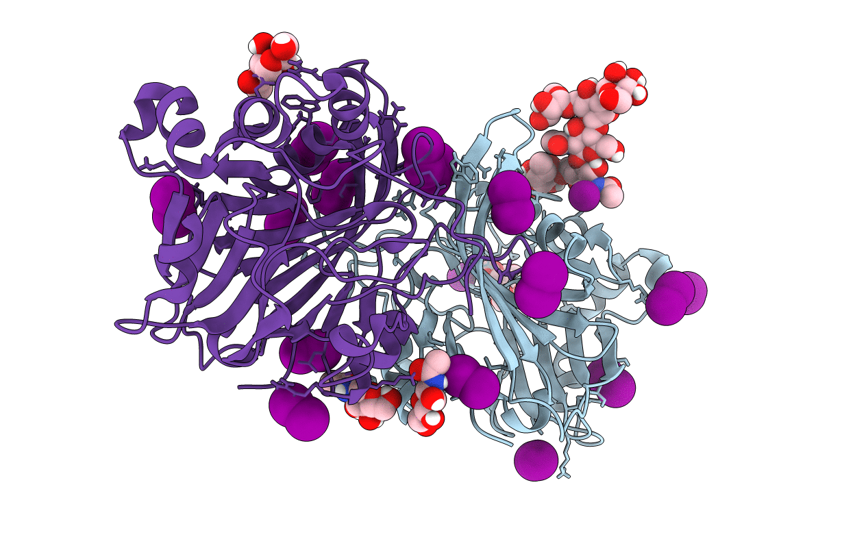
PDB ID:
6PKG
Keywords:
Title:
Zebrafish N-acetylglucosamine-1-phosphodiester alpha-N-acetylglucosaminidase (NAGPA) catalytic domain auto-inhibited by pro-peptide
Biological Source:
Source Organism(s):
Danio rerio (Taxon ID: 7955)
Expression System(s):
Method Details:
Experimental Method:
Resolution:
2.80 Å
R-Value Free:
0.20
R-Value Work:
0.18
R-Value Observed:
0.18
Space Group:
P 41 21 2
