6PB1 image
Entry Detail
PDB ID:
6PB1
EMDB ID:
Title:
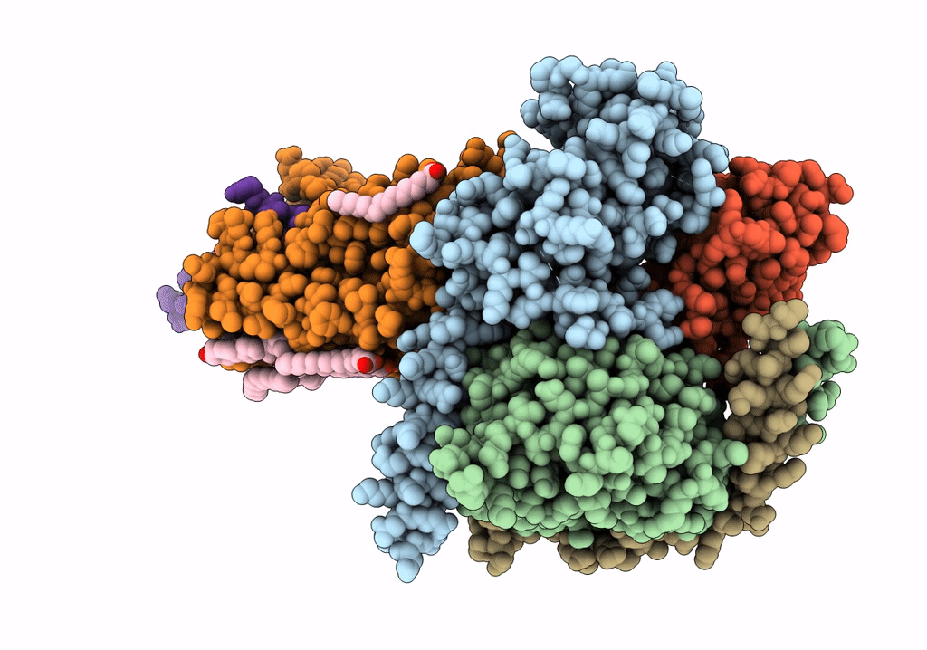
Cryo-EM structure of Urocortin 1-bound Corticotropin-releasing factor 2 receptor in complex with Gs protein and Nb35
Biological Source:
Host Organism:
PDB Version:
Deposition Date:
2019-06-12
Release Date:
2020-02-12
Method Details:
Experimental Method:
Resolution:
2.80 Å
Aggregation State:
PARTICLE
Reconstruction Method:
SINGLE PARTICLE
Macromolecular Entities
Polymer Type:polypeptide(L)
Description:Guanine nucleotide-binding protein G(s) subunit alpha isoforms short,Guanine nucleotide-binding protein G(i) subunit alpha-1,Guanine nucleotide-binding protein G(s) subunit alpha isoforms short
Mutations:A212G, S352A
Chain IDs:C (auth: A)
Chain Length:378
Number of Molecules:1
Biological Source:Homo sapiens
Polymer Type:polypeptide(L)
Description:Guanine nucleotide-binding protein G(I)/G(S)/G(T) subunit beta-1
Chain IDs:D (auth: B)
Chain Length:345
Number of Molecules:1
Biological Source:Homo sapiens
Polymer Type:polypeptide(L)
Description:Guanine nucleotide-binding protein G(I)/G(S)/G(O) subunit gamma-2
Chain IDs:E (auth: G)
Chain Length:71
Number of Molecules:1
Biological Source:Homo sapiens
Polymer Type:polypeptide(L)
Description:Nanobody 35
Chain IDs:F (auth: N)
Chain Length:140
Number of Molecules:1
Biological Source:synthetic construct
Polymer Type:polypeptide(L)
Description:Corticotropin-releasing factor receptor 2
Chain IDs:A (auth: P)
Chain Length:387
Number of Molecules:1
Biological Source:Homo sapiens
Polymer Type:polypeptide(L)
Description:Urocortin
Chain IDs:B (auth: U)
Chain Length:40
Number of Molecules:1
Biological Source:Homo sapiens
Primary Citation
Molecular Basis for Hormone Recognition and Activation of Corticotropin-Releasing Factor Receptors.
Mol.Cell 77 669 ? (2020)
PMID: 32004470 DOI: 10.1016/j.molcel.2020.01.013

Abstact

Corticotropin-releasing factor (CRF) and the three related peptides urocortins 1-3 (UCN1-UCN3) are endocrine hormones that control the stress responses by activating CRF1R and CRF2R, two members of class B G-protein-coupled receptors (GPCRs). Here, we present two cryoelectron microscopy (cryo-EM) structures of UCN1-bound CRF1R and CRF2R with the stimulatory G protein. In both structures, UCN1 adopts a single straight helix with its N terminus dipped into the receptor transmembrane bundle. Although the peptide-binding residues in CRF1R and CRF2R are different from other members of class B GPCRs, the residues involved in receptor activation and G protein coupling are conserved. In addition, both structures reveal bound cholesterol molecules to the receptor transmembrane helices. Our structures define the basis of ligand-binding specificity in the CRF receptor-hormone system, establish a common mechanism of class B GPCR activation and G protein coupling, and provide a paradigm for studying membrane protein-lipid interactions for class B GPCRs.

Legend

Protein

Chemical

Disease

Primary Citation of related structures