Deposition Date
2019-06-06
Release Date
2019-09-25
Last Version Date
2024-10-23
Entry Detail
PDB ID:
6P7S
Keywords:
Title:
Crystal Structure of the Cedar henipavirus Attachment G Glycoprotein globular domain in complex with the receptor ephrin-B1
Biological Source:
Source Organism(s):
Cedar virus (Taxon ID: 1221391)
Mus musculus (Taxon ID: 10090)
Mus musculus (Taxon ID: 10090)
Expression System(s):
Method Details:
Experimental Method:
Resolution:
3.49 Å
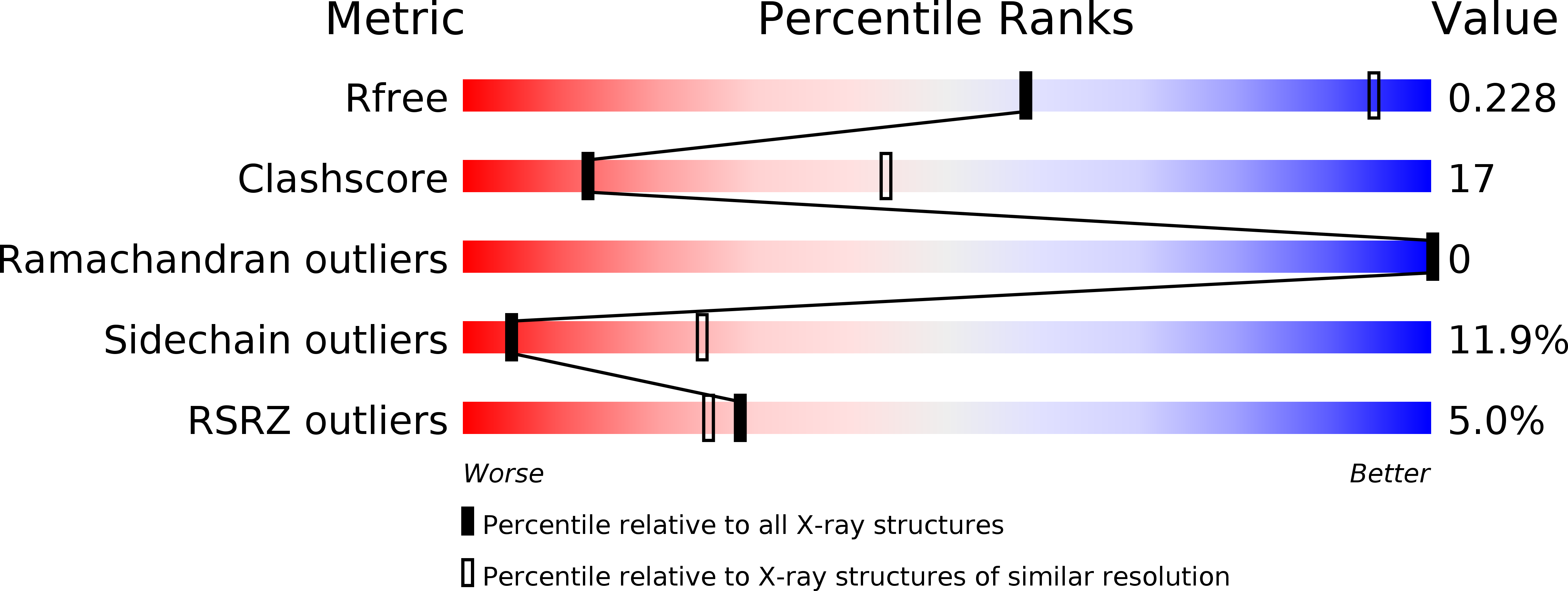
R-Value Free:
0.22
R-Value Work:
0.19
R-Value Observed:
0.19
Space Group:
P 65