6P5S image
Deposition Date 2019-05-30
Release Date 2019-07-31
Last Version Date 2024-11-13
Entry Detail
PDB ID:
6P5S
Title:
HIPK2 kinase domain bound to CX-4945
Biological Source:
Source Organism:
Homo sapiens (Taxon ID: 9606)
Host Organism:
Method Details:
Experimental Method:
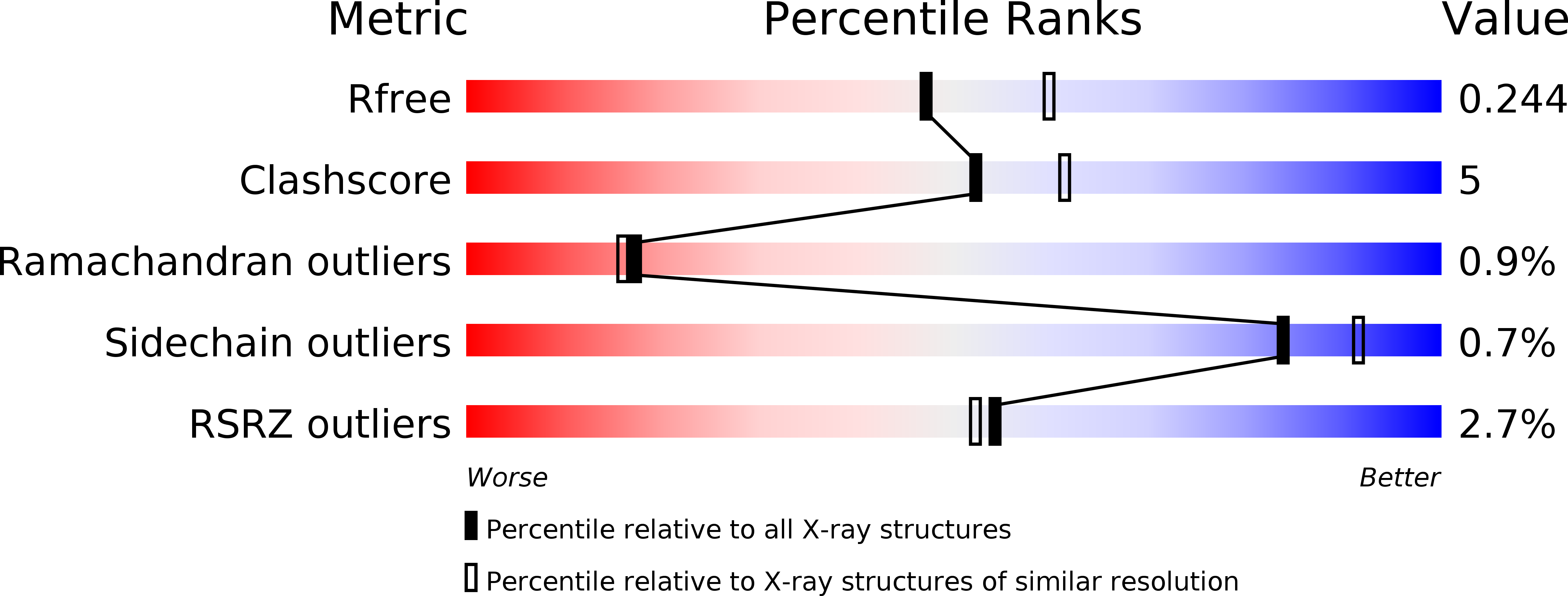
Resolution:
2.19 Å
R-Value Free:
0.23
R-Value Work:
0.19
R-Value Observed:
0.19
Space Group:
P 62
Macromolecular Entities
Polymer Type:polypeptide(L)
Molecule:Homeodomain-interacting protein kinase 2
Gene (Uniprot):HIPK2
Chain IDs:A
Chain Length:390
Number of Molecules:1
Biological Source:Homo sapiens
Modified Residue
Compound ID Chain ID Parent Comp ID Details 2D Image
PTR A TYR modified residue
SEP A SER modified residue
Ligand Molecules
Primary Citation
The crystal structure of the protein kinase HIPK2 reveals a unique architecture of its CMGC-insert region.
J.Biol.Chem. 294 13545 13559 (2019)
PMID: 31341017 DOI: 10.1074/jbc.RA119.009725

Abstact

The homeodomain-interacting protein kinase (HIPK) family is comprised of four nuclear protein kinases, HIPK1-4. HIPK proteins phosphorylate a diverse range of transcription factors involved in cell proliferation, differentiation, and apoptosis. HIPK2, thus far the best-characterized member of this largely understudied family of protein kinases, plays a role in the activation of p53 in response to DNA damage. Despite this tumor-suppressor function, HIPK2 is also found overexpressed in several cancers, and its hyperactivation causes chronic fibrosis. There are currently no structures of HIPK2 or of any other HIPK kinase. Here, we report the crystal structure of HIPK2's kinase domain bound to CX-4945, a casein kinase 2α (CK2α) inhibitor currently in clinical trials against several cancers. The structure, determined at 2.2 Å resolution, revealed that CX-4945 engages the HIPK2 active site in a hybrid binding mode between that seen in structures of CK2α and Pim1 kinases. The HIPK2 kinase domain crystallized in the active conformation, which was stabilized by phosphorylation of the activation loop. We noted that the overall kinase domain fold of HIPK2 closely resembles that of evolutionarily related dual-specificity tyrosine-regulated kinases (DYRKs). Most significant structural differences between HIPK2 and DYRKs included an absence of the regulatory N-terminal domain and a unique conformation of the CMGC-insert region and of a newly defined insert segment in the αC-β4 loop. This first crystal structure of HIPK2 paves the way for characterizing the understudied members of the HIPK family and for developing HIPK2-directed therapies for managing cancer and fibrosis.

Legend

Protein

Chemical

Disease

Primary Citation of related structures