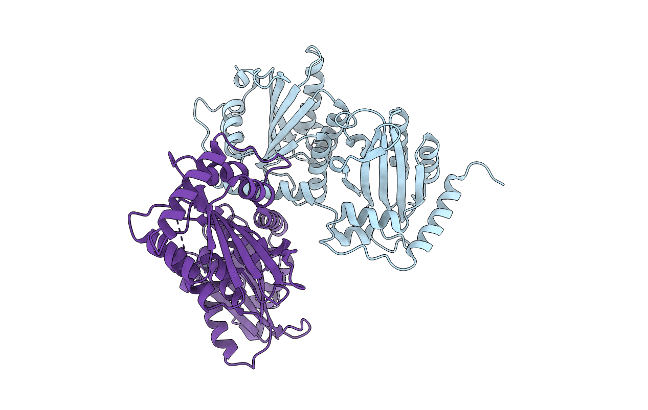
6P3Y image
Deposition Date 2019-05-25
Release Date 2020-02-12
Last Version Date 2023-08-16
Entry Detail
PDB ID:
6P3Y
Title:
Crystal Structure of Full Length APOBEC3G E/Q (pH 7.4)
Biological Source:
Source Organism:
Macaca mulatta (Taxon ID: 9544)
Host Organism:
Method Details:
Experimental Method:
Resolution:
2.57 Å
R-Value Free:
0.23
R-Value Work:
0.19
R-Value Observed:
0.19
Space Group:
P 1 21 1
Macromolecular Entities
Polymer Type:polypeptide(L)
Molecule:Apolipoprotein B mRNA editing enzyme, catalytic peptide-like 3G
Gene (Uniprot):APOBEC3G
Chain IDs:A, B
Chain Length:386
Number of Molecules:2
Biological Source:Macaca mulatta
Ligand Molecules
Primary Citation
Understanding the structural basis of HIV-1 restriction by the full length double-domain APOBEC3G.
Nat Commun 11 632 632 (2020)
PMID: 32005813 DOI: 10.1038/s41467-020-14377-y

Abstact

APOBEC3G, a member of the double-domain cytidine deaminase (CD) APOBEC, binds RNA to package into virions and restrict HIV-1 through deamination-dependent or deamination-independent inhibition. Mainly due to lack of a full-length double-domain APOBEC structure, it is unknown how CD1/CD2 domains connect and how dimerization/multimerization is linked to RNA binding and virion packaging for HIV-1 restriction. We report rhesus macaque A3G structures that show different inter-domain packing through a short linker and refolding of CD2. The A3G dimer structure has a hydrophobic dimer-interface matching with that of the previously reported CD1 structure. A3G dimerization generates a surface with intensified positive electrostatic potentials (PEP) for RNA binding and dimer stabilization. Unexpectedly, mutating the PEP surface and the hydrophobic interface of A3G does not abolish virion packaging and HIV-1 restriction. The data support a model in which only one RNA-binding mode is critical for virion packaging and restriction of HIV-1 by A3G.

Legend

Protein

Chemical

Disease

Primary Citation of related structures