6P2D image
Deposition Date 2019-05-21
Release Date 2020-06-03
Last Version Date 2025-11-12
Entry Detail
PDB ID:
6P2D
Keywords:
Title:
Structure of mouse ketohexokinase-C in complex with fructose and ADP
Biological Source:
Source Organism:
Mus musculus (Taxon ID: 10090)
Method Details:
Experimental Method:
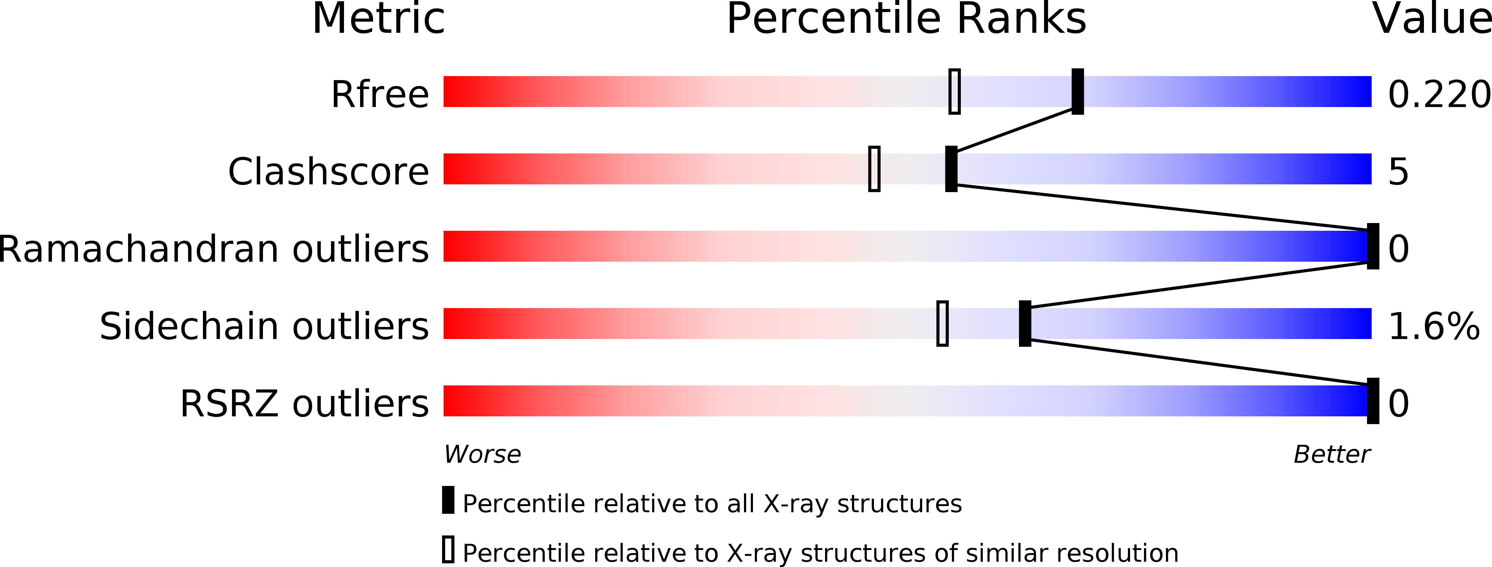
Resolution:
1.79 Å
R-Value Free:
0.22
R-Value Work:
0.17
Space Group:
C 2 2 21
Macromolecular Entities
Structures with similar UniProt ID
Protein Blast
Polymer Type:polypeptide(L)
Molecule:Ketohexokinase
Chain IDs:A
Chain Length:315
Number of Molecules:1
Biological Source:Mus musculus
Primary Citation
Michaelis-like complex of mouse ketohexokinase isoform C.
Acta Crystallogr D Struct Biol 80 377 385 (2024)
PMID: 38805243 DOI: 10.1107/S2059798324003723

Abstact

Over the past forty years there has been a drastic increase in fructose-related diseases, including obesity, heart disease and diabetes. Ketohexokinase (KHK), the first enzyme in the liver fructolysis pathway, catalyzes the ATP-dependent phosphorylation of fructose to fructose 1-phosphate. Understanding the role of KHK in disease-related processes is crucial for the management and prevention of this growing epidemic. Molecular insight into the structure-function relationship in ligand binding and catalysis by KHK is needed for the design of therapeutic inhibitory ligands. Ketohexokinase has two isoforms: ketohexokinase A (KHK-A) is produced ubiquitously at low levels, whereas ketohexokinase C (KHK-C) is found at much higher levels, specifically in the liver, kidneys and intestines. Structures of the unliganded and liganded human isoforms KHK-A and KHK-C are known, as well as structures of unliganded and inhibitor-bound mouse KHK-C (mKHK-C), which shares 90% sequence identity with human KHK-C. Here, a high-resolution X-ray crystal structure of mKHK-C refined to 1.79 Å resolution is presented. The structure was determined in a complex with both the substrate fructose and the product of catalysis, ADP, providing a view of the Michaelis-like complex of the mouse ortholog. Comparison to unliganded structures suggests that KHK undergoes a conformational change upon binding of substrates that places the enzyme in a catalytically competent form in which the β-sheet domain from one subunit rotates by 16.2°, acting as a lid for the opposing active site. Similar kinetic parameters were calculated for the mouse and human enzymes and indicate that mice may be a suitable animal model for the study of fructose-related diseases. Knowledge of the similarity between the mouse and human enzymes is important for understanding preclinical efforts towards targeting this enzyme, and this ground-state, Michaelis-like complex suggests that a conformational change plays a role in the catalytic function of KHK-C.

Legend

Protein

Chemical

Disease

Primary Citation of related structures
Feedback Form
Name
Email
Institute
Feedback