6P0T image
Deposition Date 2019-05-17
Release Date 2019-06-19
Last Version Date 2023-10-11
Entry Detail
PDB ID:
6P0T
Title:
Crystal structure of ternary DNA complex "FX(1-2)-1Xis" containing E. coli Fis and phage lambda Xis
Biological Source:
Source Organism:
Method Details:
Experimental Method:
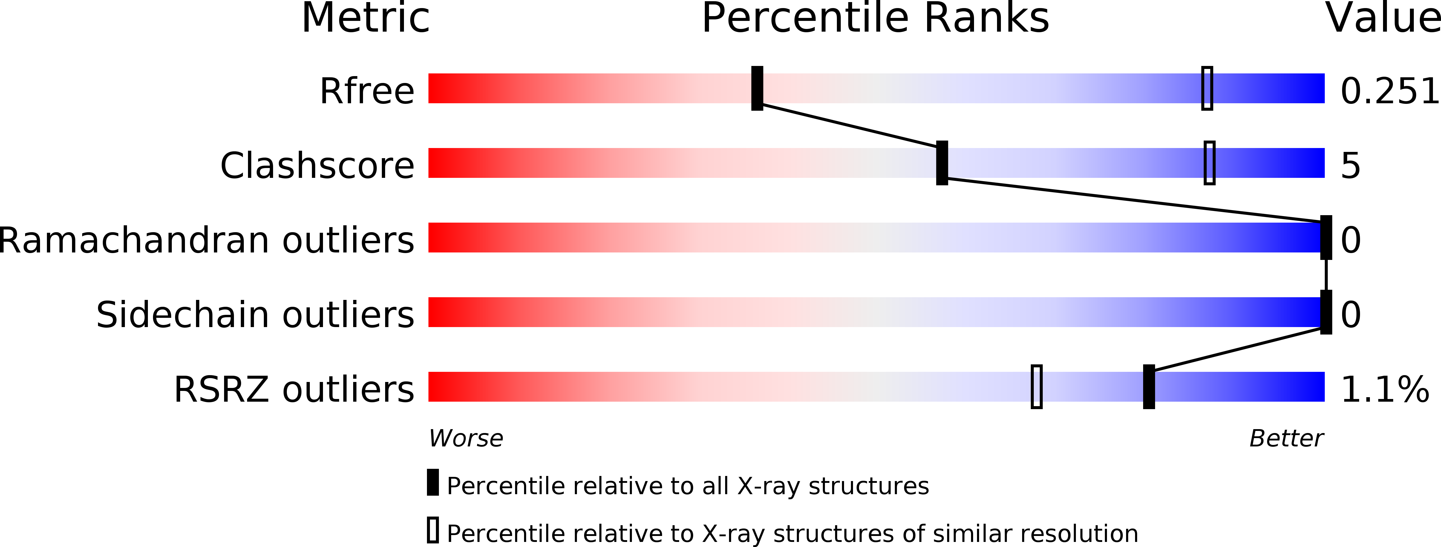
Resolution:
3.60 Å
R-Value Free:
0.24
R-Value Work:
0.19
R-Value Observed:
0.20
Space Group:
P 43 21 2
Macromolecular Entities
Structures with similar UniProt ID
Protein Blast
Polymer Type:polypeptide(L)
Molecule:DNA-binding protein Fis
Gene (Uniprot):fis
Chain IDs:A (auth: B), B (auth: A)
Chain Length:98
Number of Molecules:2
Biological Source:Escherichia coli
Polymer Type:polydeoxyribonucleotide
Molecule:DNA (27-MER), FX1-2
Chain IDs:C
Chain Length:27
Number of Molecules:1
Biological Source:Escherichia coli
Polymer Type:polydeoxyribonucleotide
Molecule:DNA (27-MER), FX1-2
Chain IDs:D
Chain Length:27
Number of Molecules:1
Biological Source:Escherichia coli
Structures with similar UniProt ID
Protein Blast
Polymer Type:polypeptide(L)
Molecule:Excisionase
Gene (Uniprot):xis
Chain IDs:E
Chain Length:55
Number of Molecules:1
Biological Source:Escherichia phage lambda
Ligand Molecules
Primary Citation
Cooperative DNA binding by proteins through DNA shape complementarity.
Nucleic Acids Res. 47 8874 8887 (2019)
PMID: 31616952 DOI: 10.1093/nar/gkz642

Abstact

Localized arrays of proteins cooperatively assemble onto chromosomes to control DNA activity in many contexts. Binding cooperativity is often mediated by specific protein-protein interactions, but cooperativity through DNA structure is becoming increasingly recognized as an additional mechanism. During the site-specific DNA recombination reaction that excises phage λ from the chromosome, the bacterial DNA architectural protein Fis recruits multiple λ-encoded Xis proteins to the attR recombination site. Here, we report X-ray crystal structures of DNA complexes containing Fis + Xis, which show little, if any, contacts between the two proteins. Comparisons with structures of DNA complexes containing only Fis or Xis, together with mutant protein and DNA binding studies, support a mechanism for cooperative protein binding solely by DNA allostery. Fis binding both molds the minor groove to potentiate insertion of the Xis β-hairpin wing motif and bends the DNA to facilitate Xis-DNA contacts within the major groove. The Fis-structured minor groove shape that is optimized for Xis binding requires a precisely positioned pyrimidine-purine base-pair step, whose location has been shown to modulate minor groove widths in Fis-bound complexes to different DNA targets.

Legend

Protein

Chemical

Disease

Primary Citation of related structures