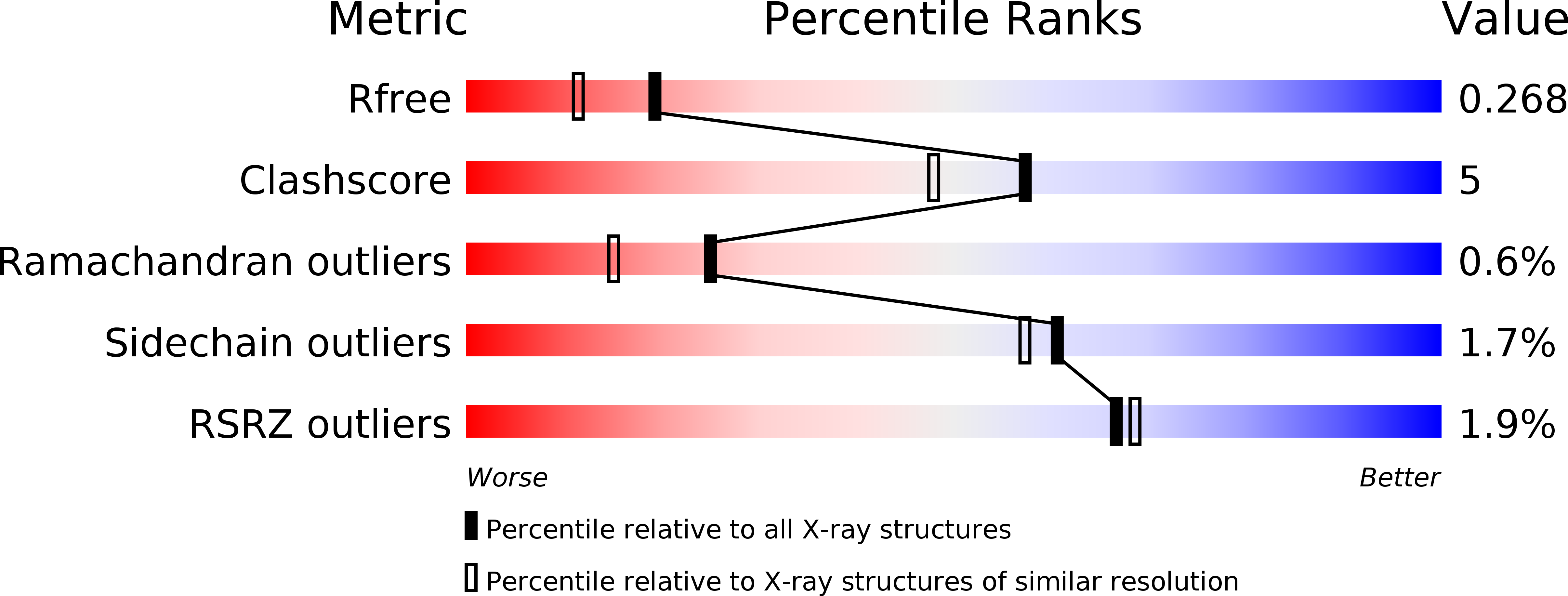
6OZW image
Deposition Date 2019-05-16
Release Date 2019-06-26
Last Version Date 2023-10-11
Entry Detail
PDB ID:
6OZW
Title:
Crystal structure of the 65-kilodalton amino-terminal fragment of DNA topoisomerase I from Streptococcus mutans
Biological Source:
Method Details:
Experimental Method:
Resolution:
2.06 Å
R-Value Free:
0.26
R-Value Work:
0.20
R-Value Observed:
0.20
Space Group:
P 21 21 2
Macromolecular Entities
Structures with similar UniProt ID
Protein Blast
Polymer Type:polypeptide(L)
Molecule:DNA topoisomerase 1
Gene (Uniprot):topA
Chain IDs:A
Chain Length:595
Number of Molecules:1
Biological Source:Streptococcus mutans serotype c (strain ATCC 700610 / UA159)
Primary Citation
Crystal structure of the 65-kilodalton amino-terminal fragment of DNA topoisomerase I from the gram-positive model organism Streptococcus mutans.
Biochem.Biophys.Res.Commun. 516 333 338 (2019)
PMID: 31204053 DOI: 10.1016/j.bbrc.2019.06.034

Abstact

Herein we report the first structure of topoisomerase I determined from the gram-positive bacterium, S. mutans. Bacterial topoisomerase I is an ATP-independent type 1A topoisomerase that uses the inherent torsional strain within hyper-negatively supercoiled DNA as an energy source for its critical function of DNA relaxation. Interest in the enzyme has gained momentum as it has proven to be essential in various bacterial organisms. In order to aid in further biochemical characterization, the apo 65-kDa amino-terminal fragment of DNA topoisomerase I from the gram-positive model organism Streptococcus mutans was crystalized and a three-dimensional structure was determined to 2.06 Å resolution via x-ray crystallography. The overall structure illustrates the four classic major domains that create the traditional topoisomerase I "lock" formation comprised of a sizable toroidal aperture atop what is considered to be a highly dynamic body. A catalytic tyrosine residue resides at the interface between two domains and is known to form a 5' phosphotyrosine DNA-enzyme intermediate during transient single-stranded cleavage required for enzymatic relaxation of hyper negative DNA supercoils. Surrounding the catalytic tyrosine residue is the remainder of the highly conserved active site. Within 5 Å from the catalytic center, only one dissimilar residue is observed between topoisomerase I from S. mutans and the gram-negative model organism E. coli. Immediately adjacent to the conserved active site, however, S. mutans topoisomerase I displays a somewhat unique nine residue loop extension not present in any bacterial topoisomerase I structures previously determined other than that of an extremophile.

Legend

Protein

Chemical

Disease

Primary Citation of related structures