
Deposition Date
2019-05-16
Release Date
2019-12-11
Last Version Date
2024-05-15
Entry Detail
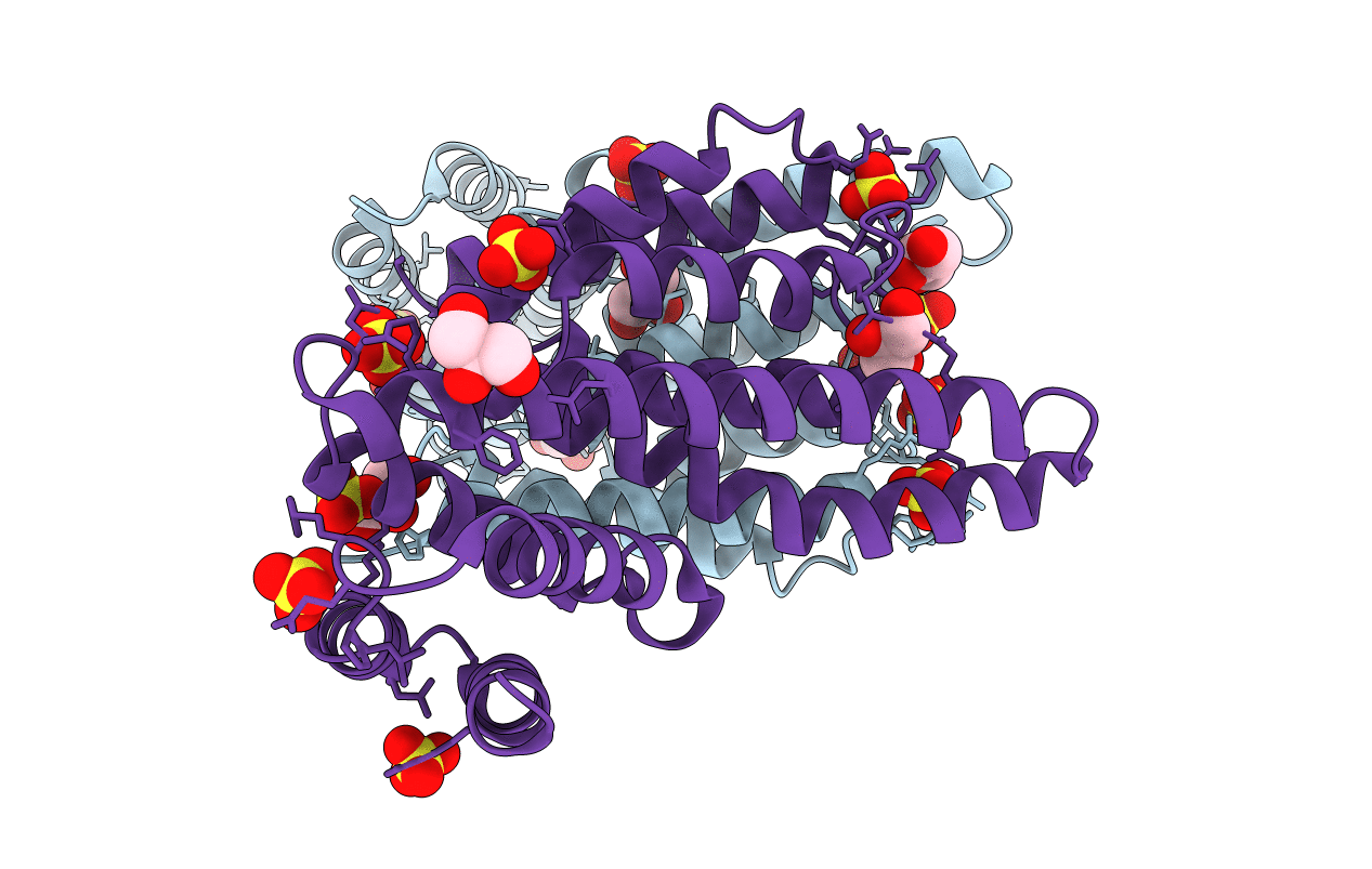
PDB ID:
6OZU
Keywords:
Title:
Crystal structure of the MIF4G domain of Trypanosoma cruzi translation initiation factor EIF4G5
Biological Source:
Source Organism:
Trypanosoma cruzi (Taxon ID: 5693)
Host Organism:
Method Details:
Experimental Method:
Resolution:
2.40 Å
R-Value Free:
0.24
R-Value Work:
0.20
R-Value Observed:
0.20
Space Group:
P 41 21 2
