6OZE image
Deposition Date 2019-05-15
Release Date 2019-09-04
Last Version Date 2023-10-11
Entry Detail
PDB ID:
6OZE
Keywords:
Title:
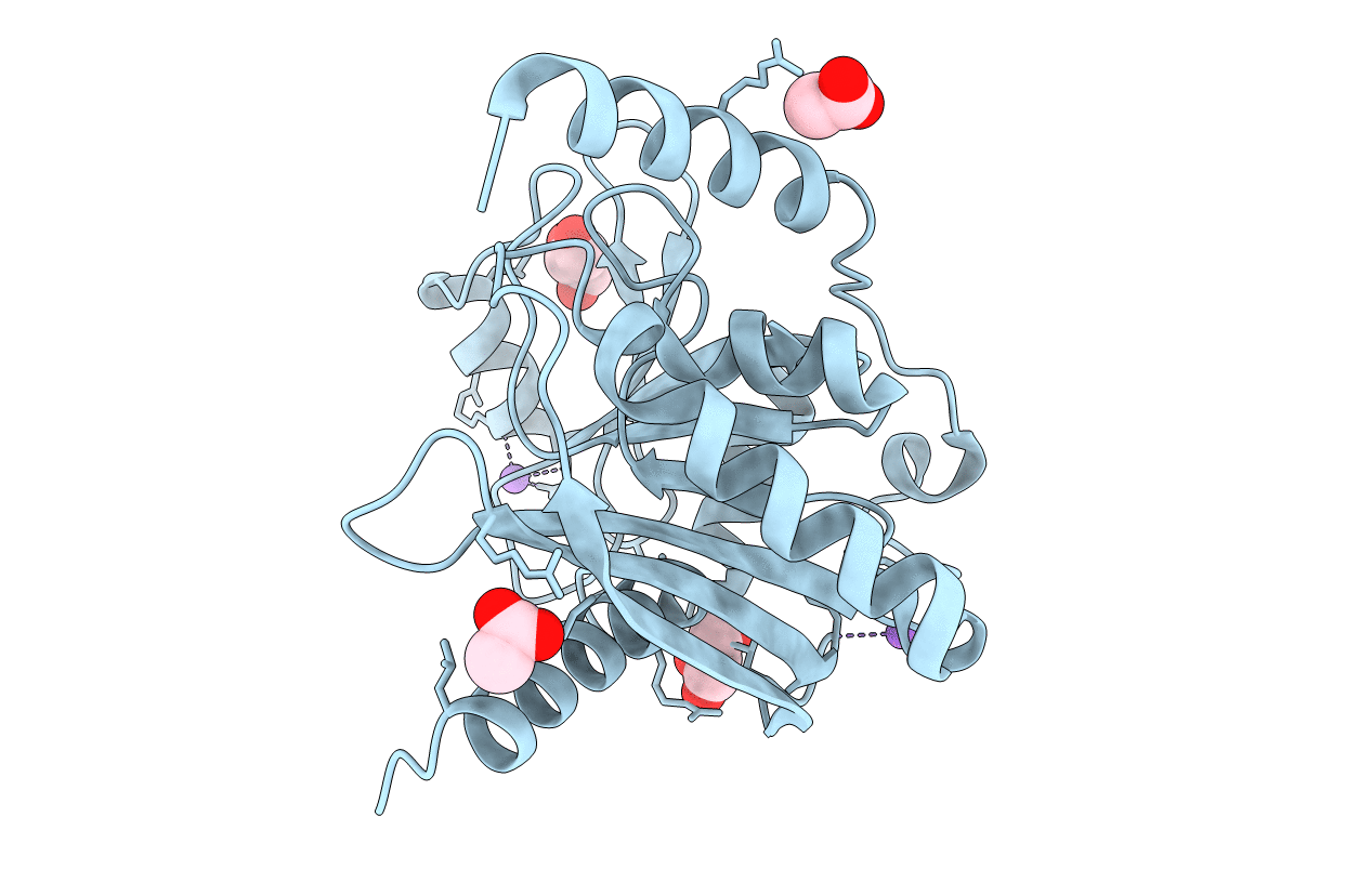
Crystal structure of the catalytic domain of human Endonuclease V (C140S/C225S/C226A/C228S)
Biological Source:
Source Organism:
Homo sapiens (Taxon ID: 9606)
Host Organism:
Method Details:
Experimental Method:
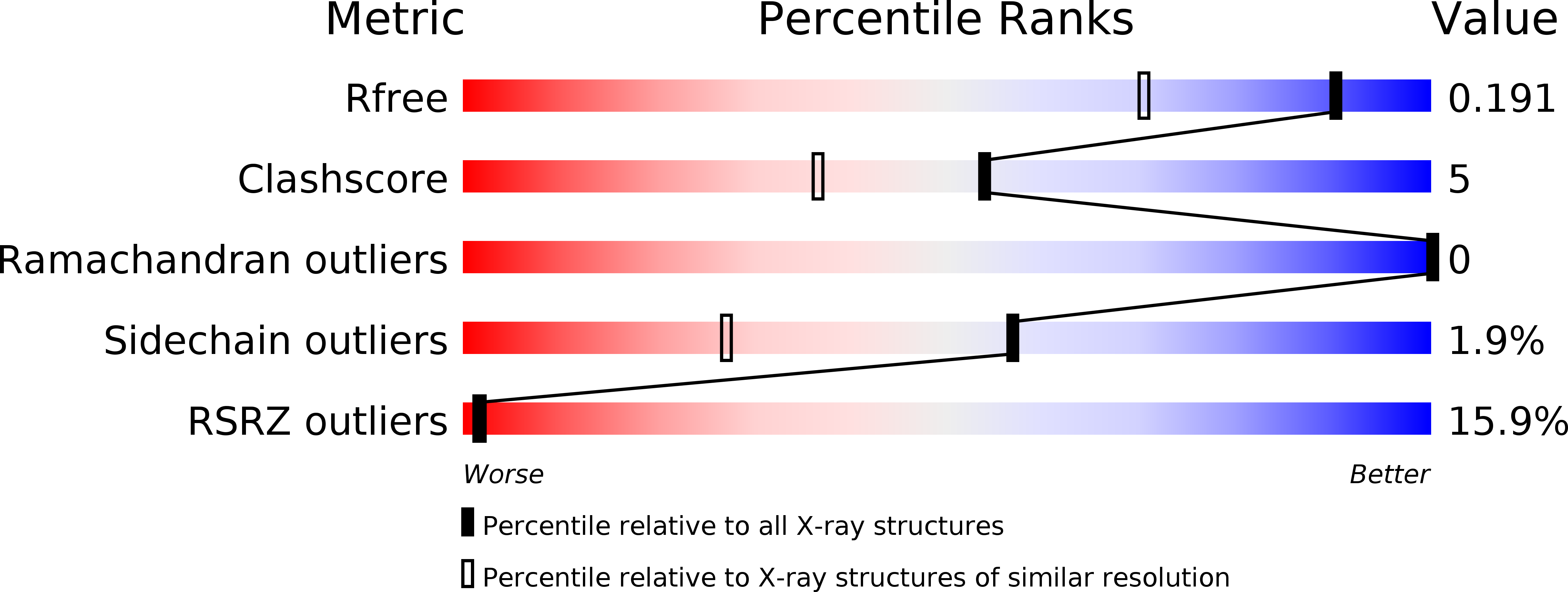
Resolution:
1.50 Å
R-Value Free:
0.19
R-Value Work:
0.17
R-Value Observed:
0.17
Space Group:
P 31
Macromolecular Entities
Structures with similar UniProt ID
Protein Blast
Polymer Type:polypeptide(L)
Molecule:Endonuclease V
Gene (Uniprot):ENDOV
Mutagens:C140S/C225S/C226A/C228S
Chain IDs:A
Chain Length:246
Number of Molecules:1
Biological Source:Homo sapiens
Primary Citation
Evolution of Inosine-Specific Endonuclease V from Bacterial DNase to Eukaryotic RNase.
Mol.Cell 76 44 ? (2019)
PMID: 31444105 DOI: 10.1016/j.molcel.2019.06.046

Abstact

Endonuclease V (EndoV) cleaves the second phosphodiester bond 3' to a deaminated adenosine (inosine). Although highly conserved, EndoV homologs change substrate preference from DNA in bacteria to RNA in eukaryotes. We have characterized EndoV from six different species and determined crystal structures of human EndoV and three EndoV homologs from bacteria to mouse in complex with inosine-containing DNA/RNA hybrid or double-stranded RNA (dsRNA). Inosine recognition is conserved, but changes in several connecting loops in eukaryotic EndoV confer recognition of 3 ribonucleotides upstream and 7 or 8 bp of dsRNA downstream of the cleavage site, and bacterial EndoV binds only 2 or 3 nt flanking the scissile phosphate. In addition to the two canonical metal ions in the active site, a third Mn2+ that coordinates the nucleophilic water appears necessary for product formation. Comparison of EndoV with its homologs RNase H1 and Argonaute reveals the principles by which these enzymes recognize RNA versus DNA.

Legend

Protein

Chemical

Disease

Primary Citation of related structures