6OTX image
Deposition Date 2019-05-03
Release Date 2020-04-01
Last Version Date 2023-10-11
Entry Detail
PDB ID:
6OTX
Title:
Crystallographic Structure of (HbII-HbIII)-O2 from Lucina pectinata at pH 7.0
Biological Source:
Source Organism:
Host Organism:
Method Details:
Experimental Method:
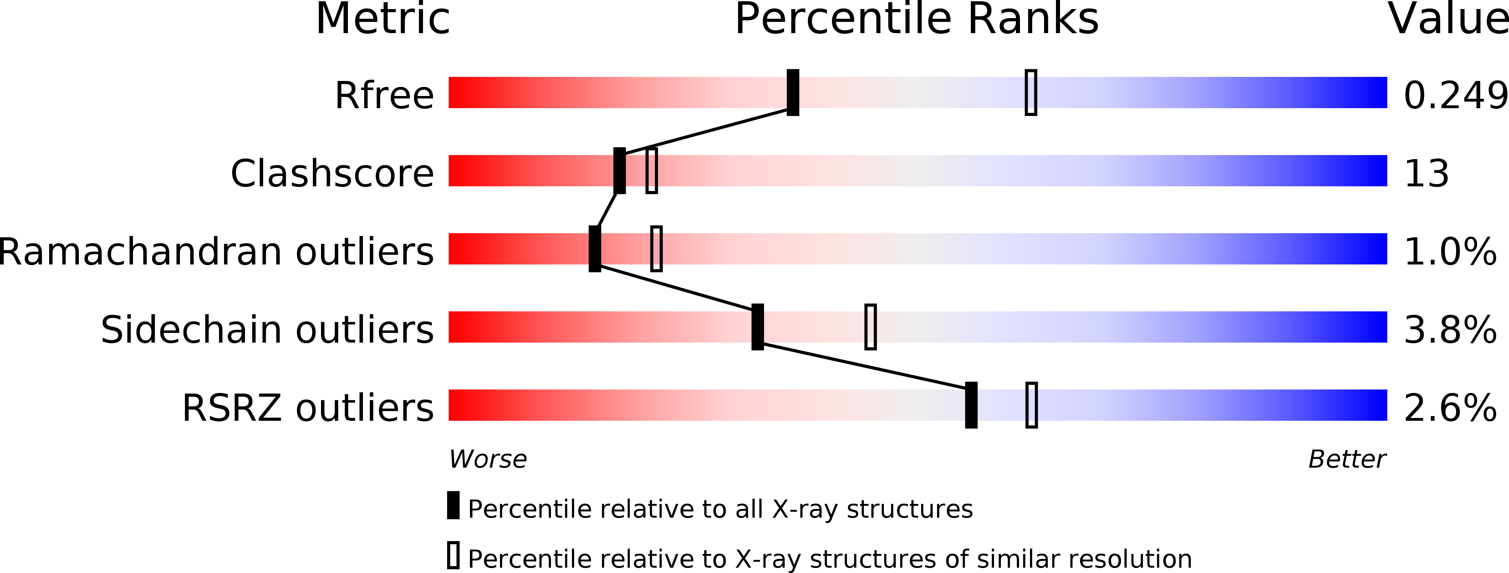
Resolution:
2.54 Å
R-Value Free:
0.24
R-Value Work:
0.19
R-Value Observed:
0.19
Space Group:
P 42 21 2
Macromolecular Entities
Structures with similar UniProt ID
Protein Blast
Polymer Type:polypeptide(L)
Molecule:Hemoglobin II
Chain IDs:A
Chain Length:152
Number of Molecules:1
Biological Source:Phacoides pectinatus
Structures with similar UniProt ID
Protein Blast
Polymer Type:polypeptide(L)
Molecule:Hemoglobin III
Chain IDs:B
Chain Length:152
Number of Molecules:1
Biological Source:Phacoides pectinatus
Primary Citation
Lucina pectinata oxyhemoglobin (II-III) heterodimer pH susceptibility.
J.Inorg.Biochem. 207 111055 111055 (2020)
PMID: 32217352 DOI: 10.1016/j.jinorgbio.2020.111055

Abstact

Lucina pectinata live in high concentrations of hydrogen sulfide (H2S) and contains one hemoglobin, Hemoglobin I (HbI), transporting H2S and two hemoglobins, Hemoglobin II (HbII) and Hemoglobin (HbIII), transferring dioxygen to symbionts. HbII and HbIII contain B10 tyrosine (Tyr) and E7 glutamine (Gln) in the heme pocket generating an efficient hydrogen bonding network with the (HbII-HbIII)-O2 species, leading to very low ligand dissociation rates. The results indicate that the oxy-hemeprotein is susceptible to pH from 4 to 9, at acidic conditions, and as a function of the potassium ferricyanide concentration, 100% of the met-aquo derivative is produced. Without a strong oxidant, pH 5 generates a small concentration of the met-aquo complex. The process is accelerated by the presence of salts, as indicated by the crystallization structures and UV-Vis spectra. The results suggest that acidic pH generates conformational changes associated with B10 and E7 heme pocket amino acids, weakening the (HbII-HbIII)-O2 hydrogen bond network. The observation is supported by X-ray crystallography, since at pH 4 and 5, the heme-Fe tends to oxidize, while at pH 7, the oxy-heterodimer is present. Conformational changes also are observed at higher pH by the presence of a 605 nm transition associated with the iron heme-Tyr interaction. Therefore, pH is one crucial factor regulating the (HbII-HbIII)-O2 complex hydrogen-bonding network. Thus, it can be proposed that the hydrogen bonding adjustments between the heme bound O2 and the Tyr and Gln amino acids contribute to oxygen dissociation from the (HbII-HbIII)-O2 system.

Legend

Protein

Chemical

Disease

Primary Citation of related structures
Feedback Form
Name
Email
Institute
Feedback