6OM8 image
Entry Detail
PDB ID:
6OM8
Title:
Caenorhabditis Elegans UDP-Glucose Dehydrogenase in complex with UDP-Xylose
Biological Source:
Source Organism:
Host Organism:
PDB Version:
Deposition Date:
2019-04-18
Release Date:
2019-05-22
Method Details:
Experimental Method:
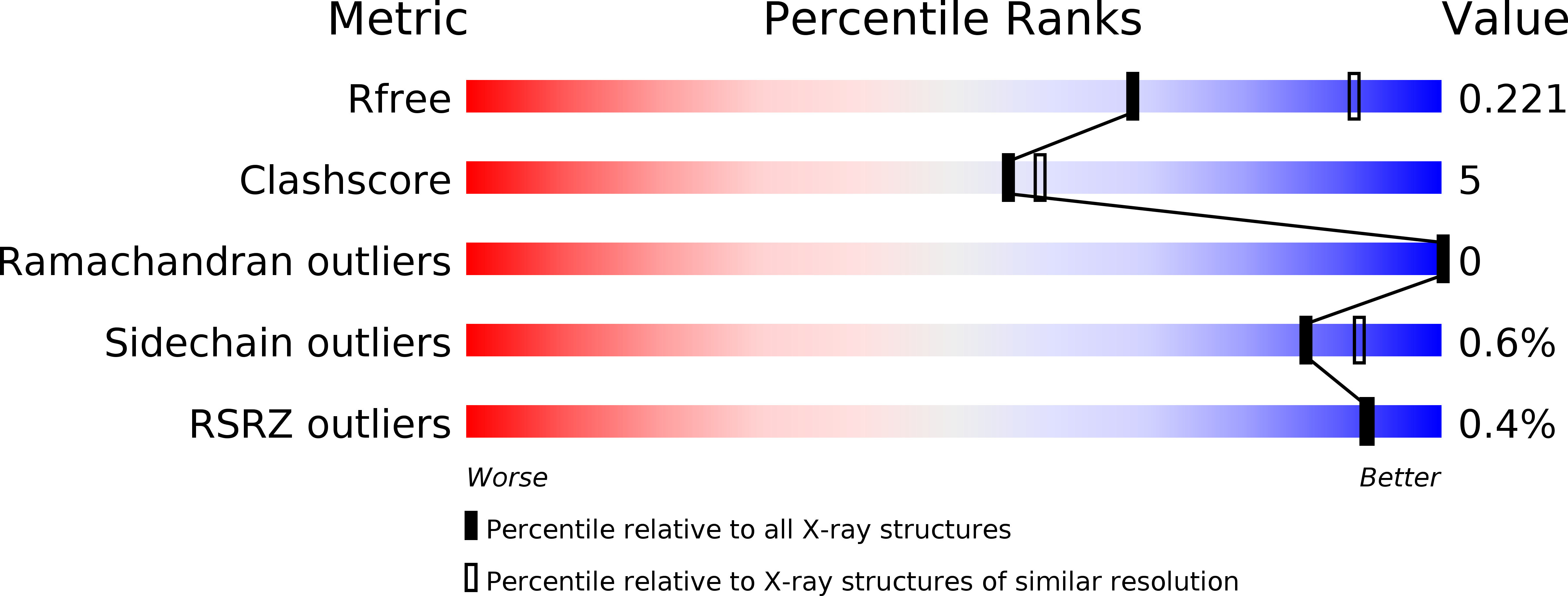
Resolution:
2.45 Å
R-Value Free:
0.22
R-Value Work:
0.19
R-Value Observed:
0.19
Space Group:
P 2 21 21
Macromolecular Entities
Polymer Type:polypeptide(L)
Description:UDP-glucose 6-dehydrogenase
Chain IDs:A, B, C, D, E, F, G, H, I, J, K, L
Chain Length:481
Number of Molecules:12
Biological Source:Caenorhabditis elegans
Ligand Molecules
Primary Citation
Conservation of Atypical Allostery inC. elegansUDP-Glucose Dehydrogenase.
Acs Omega 4 16318 16329 (2019)
PMID: 31616809 DOI: 10.1021/acsomega.9b01565

Abstact

Human UDP-glucose dehydrogenase (hUGDH) oxidizes uridine diphosphate (UDP)-glucose to UDP-glucuronic acid, an essential substrate in the phase II metabolism of drugs. The activity of hUGDH is controlled by an atypical allosteric mechanism in which the feedback inhibitor UDP-xylose competes with the substrate for the active site and triggers a buried allosteric switch to produce an inactive complex (EΩ). Previous comparisons with a nonallosteric UGDH identified six large-to-small substitutions that produce packing defects in the protein core and provide the conformational flexibility necessary for the allosteric transition. Here, we test the hypothesis that these large-to-small substitutions form a motif that can be used to identify allosteric UGDHs. Caenorhabditis elegans UGDH (cUGDH) conserves this motif with the exception of an Ala-to-Pro substitution in position 109. The crystal structures of unliganded and UDP-xylose bound cUGDH show that the A109P substitution is accommodated by an Asn-to-Ser substitution at position 290. Steady-state analysis and sedimentation velocity studies show that the allosteric transition is conserved in cUGDH. The enzyme also exhibits hysteresis in progress curves and negative cooperativity with respect to NAD+ binding. Both of these phenomena are conserved in the human enzyme, which is strong evidence that these represent fundamental features of atypical allostery in UGDH. A phylogenetic analysis of UGDH shows that the atypical allostery motif is ancient and identifies a potential transition point in the evolution of the UGDH family.

Legend

Protein

Chemical

Disease

Primary Citation of related structures