
Deposition Date
2019-04-16
Release Date
2019-12-11
Last Version Date
2024-11-20
Entry Detail
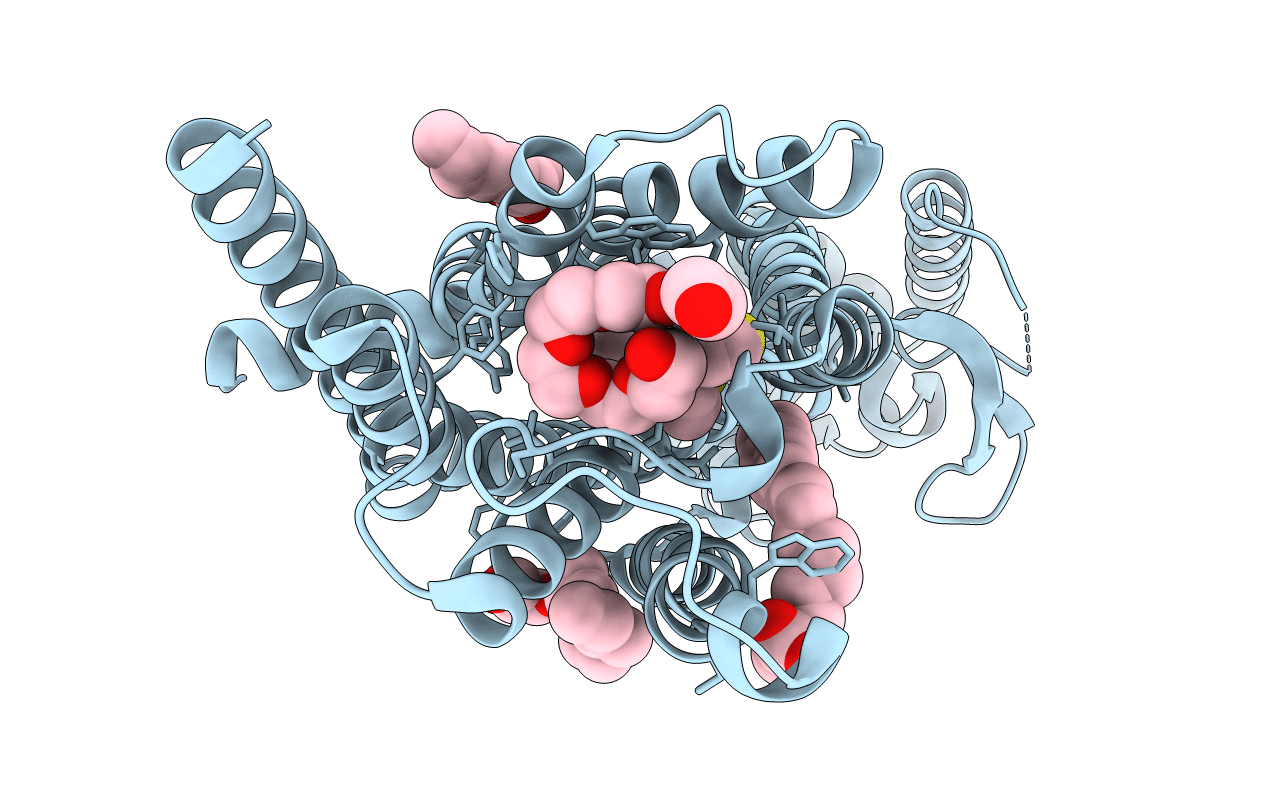
PDB ID:
6OL9
Keywords:
Title:
Structure of the M5 muscarinic acetylcholine receptor (M5-T4L) bound to tiotropium
Biological Source:
Source Organism(s):
Homo sapiens (Taxon ID: 9606)
Enterobacteria phage T4 (Taxon ID: 10665)
Enterobacteria phage T4 (Taxon ID: 10665)
Expression System(s):
Method Details:
Experimental Method:
Resolution:
2.54 Å
R-Value Free:
0.25
R-Value Work:
0.23
R-Value Observed:
0.23
Space Group:
C 1 2 1
