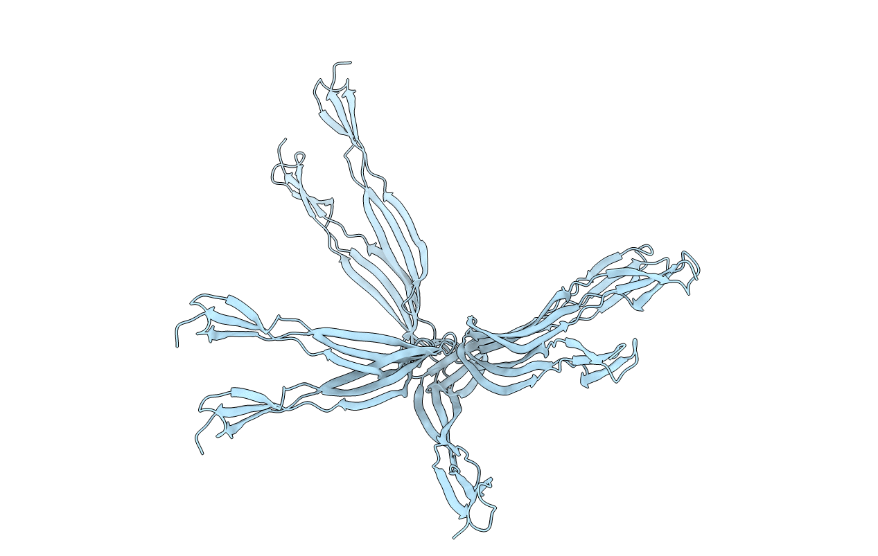
6OH1 image
Entry Detail
PDB ID:
6OH1
Keywords:
Title:
IgA1 Protease G5 domain structure
Biological Source:
Source Organism:
Host Organism:
PDB Version:
Deposition Date:
2019-04-04
Release Date:
2020-02-26
Method Details:
Experimental Method:
Conformers Calculated:
8
Conformers Submitted:
8
Selection Criteria:
structures with the lowest energy
Macromolecular Entities
Polymer Type:polypeptide(L)
Description:Immunoglobulin A1 protease
Chain IDs:A
Chain Length:84
Number of Molecules:1
Biological Source:Streptococcus pneumoniae
Ligand Molecules
Primary Citation
Streptococcus pneumoniae G5 domains bind different ligands.
Protein Sci. 28 1797 1805 (2019)
PMID: 31390088 DOI: 10.1002/pro.3693

Abstact

Many bacterial pathogens express small G5 domains that exist in the context of various membrane-anchored proteins and these G5 domains have been associated with colonization, cellular adhesion, and biofilm formation. However, despite over a decade since the computational prediction of these G5 domains, many remain uncharacterized, particularly those from Streptococcus pneumoniae. Of five previously predicted G5 domains we found that four of these, all derived from S. pneumoniae, are independently folded modules. As one of these exhibits extreme line broadening due to self-association, we were able to use NMR solution studies to probe the potential ligand interactions of the remaining three G5 domains. None of these G5 domains engage N-acetylglucosamine (NAG) as previously predicted but do interact with other small molecules that may modulate adherence to both bacteria and host cells. Specifically, while all G5 domains tested engage Zn, only one of these G5 domains engage heparin. NMR solution structural studies of the IgA1 Protease G5 (IgA1P-G5) and endo-beta-N-acetylglucosaminidase-D G5 (ENDD-G5) also facilitated identification of the ligand binding sites and confirm the typical G5 fold that comprises two connected β-sheets with no canonical core. NMR relaxation experiments indicate flexibility on both ends and within the connecting regions between the β-sheets. Our studies thus establish a basis for future biological experiments to test whether the ligands presented here are involved in bacterial adherence, either to bacteria or to host cells.

Legend

Protein

Chemical

Disease

Primary Citation of related structures