6OFC image
Deposition Date 2019-03-28
Release Date 2020-01-08
Last Version Date 2023-10-25
Entry Detail
PDB ID:
6OFC
Keywords:
Title:
Crystal structure of M. tuberculosis glutamine-dependent NAD+ synthetase complexed with Sulfonamide derivative 1, pyrophosphate, and glutamine
Biological Source:
Source Organism:
Method Details:
Experimental Method:
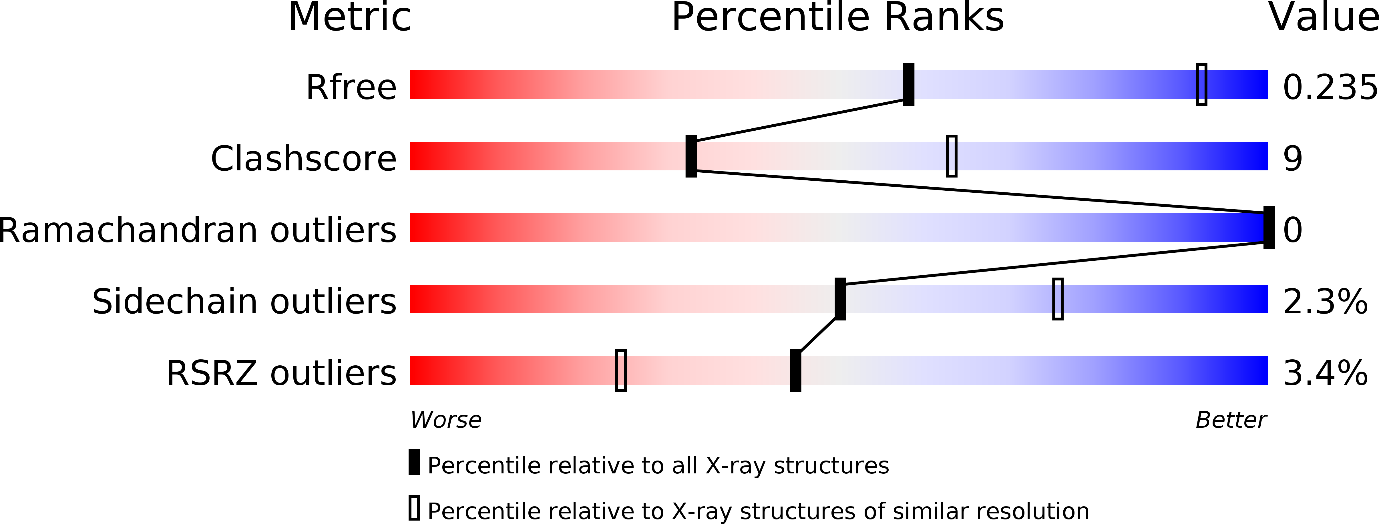
Resolution:
3.14 Å
R-Value Free:
0.23
R-Value Work:
0.18
R-Value Observed:
0.18
Space Group:
P 41 21 2
Macromolecular Entities
Structures with similar UniProt ID
Protein Blast
Polymer Type:polypeptide(L)
Molecule:Glutamine-dependent NAD(+) synthetase
Mutagens:C176A
Chain IDs:A, B, C, D
Chain Length:679
Number of Molecules:4
Biological Source:Mycobacterium tuberculosis CDC1551
Primary Citation
Different ways to transport ammonia in human and Mycobacterium tuberculosis NAD+synthetases.
Nat Commun 11 16 16 (2020)
PMID: 31911602 DOI: 10.1038/s41467-019-13845-4

Abstact

NAD+ synthetase is an essential enzyme of de novo and recycling pathways of NAD+ biosynthesis in Mycobacterium tuberculosis but not in humans. This bifunctional enzyme couples the NAD+ synthetase and glutaminase activities through an ammonia tunnel but free ammonia is also a substrate. Here we show that the Homo sapiens NAD+ synthetase (hsNadE) lacks substrate specificity for glutamine over ammonia and displays a modest activation of the glutaminase domain compared to tbNadE. We report the crystal structures of hsNadE and NAD+ synthetase from M. tuberculosis (tbNadE) with synthetase intermediate analogues. Based on the observed exclusive arrangements of the domains and of the intra- or inter-subunit tunnels we propose a model for the inter-domain communication mechanism for the regulation of glutamine-dependent activity and NH3 transport. The structural and mechanistic comparison herein reported between hsNadE and tbNadE provides also a starting point for future efforts in the development of anti-TB drugs.

Legend

Protein

Chemical

Disease

Primary Citation of related structures