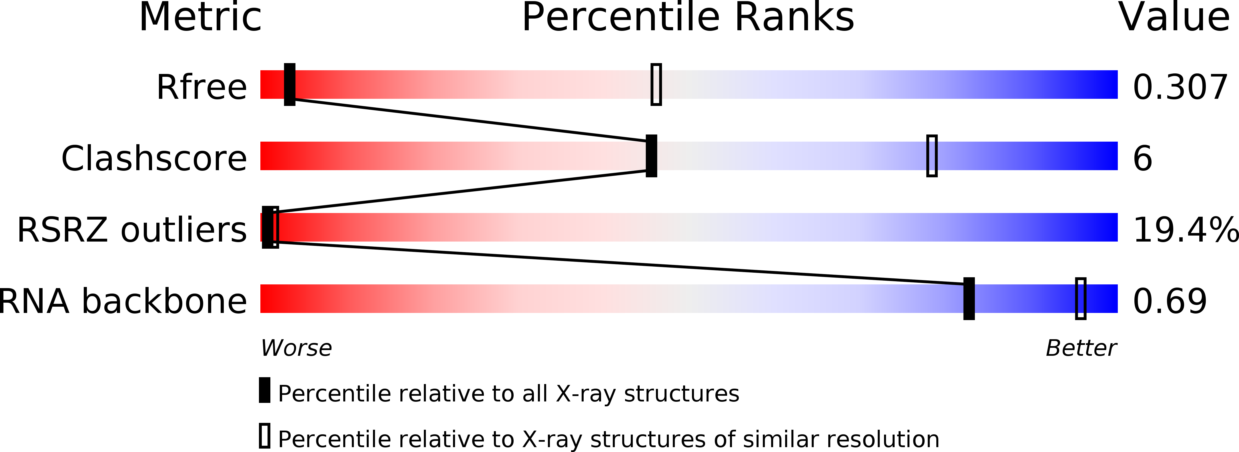
6OD9 image
Entry Detail
PDB ID:
6OD9
Keywords:
Title:
Co-crystal structure of the Fusobacterium ulcerans ZTP riboswitch using an X-ray free-electron laser
Biological Source:
Source Organism:
PDB Version:
Deposition Date:
2019-03-26
Release Date:
2019-07-17
Method Details:
Experimental Method:
Resolution:
4.10 Å
R-Value Free:
0.30
R-Value Work:
0.25
R-Value Observed:
0.26
Space Group:
P 32 2 1
Macromolecular Entities
Polymer Type:polyribonucleotide
Description:RNA (75-MER)
Chain IDs:A, B
Chain Length:75
Number of Molecules:2
Biological Source:Fusobacterium ulcerans
Primary Citation
Co-crystal structure of the Fusobacterium ulcerans ZTP riboswitch using an X-ray free-electron laser.
Acta Crystallogr.,Sect.F 75 496 500 (2019)
PMID: 31282869 DOI: 10.1107/S2053230X19008549

Abstact

Riboswitches are conformationally dynamic RNAs that regulate gene expression by binding specific small molecules. ZTP riboswitches bind the purine-biosynthetic intermediate 5-aminoimidazole-4-carboxamide riboside 5'-monophosphate (ZMP) and its triphosphorylated form (ZTP). Ligand binding to this riboswitch ultimately upregulates genes involved in folate and purine metabolism. Using an X-ray free-electron laser (XFEL), the room-temperature structure of the Fusobacterium ulcerans ZTP riboswitch bound to ZMP has now been determined at 4.1 Å resolution. This model, which was refined against a data set from ∼750 diffraction images (each from a single crystal), was found to be consistent with that previously obtained from data collected at 100 K using conventional synchrotron X-radiation. These experiments demonstrate the feasibility of time-resolved XFEL experiments to understand how the ZTP riboswitch accommodates cognate ligand binding.

Legend

Protein

Chemical

Disease

Primary Citation of related structures