Deposition Date
2019-03-21
Release Date
2019-06-05
Last Version Date
2024-03-13
Entry Detail
PDB ID:
6OBV
Keywords:
Title:
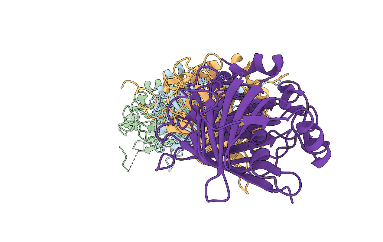
Structural insights into dehydratase substrate selection for the borrelidin and fluvirucin polyketide synthases
Biological Source:
Source Organism(s):
Actinomadura vulgaris (Taxon ID: 1233071)
Expression System(s):
Method Details:
Experimental Method:
Resolution:
2.01 Å
R-Value Free:
0.26
R-Value Work:
0.19
R-Value Observed:
0.19
Space Group:
C 1 2 1