Deposition Date
2019-03-17
Release Date
2020-04-22
Last Version Date
2024-11-20
Entry Detail
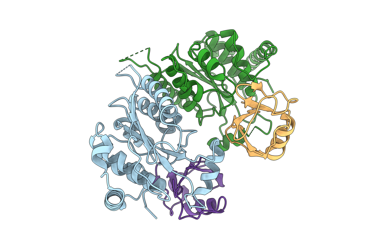
Biological Source:
Source Organism(s):
Chlamydia trachomatis serovar L2 (strain 434/Bu / ATCC VR-902B) (Taxon ID: 471472)
Homo sapiens (Taxon ID: 9606)
Homo sapiens (Taxon ID: 9606)
Expression System(s):
Method Details:
Experimental Method:
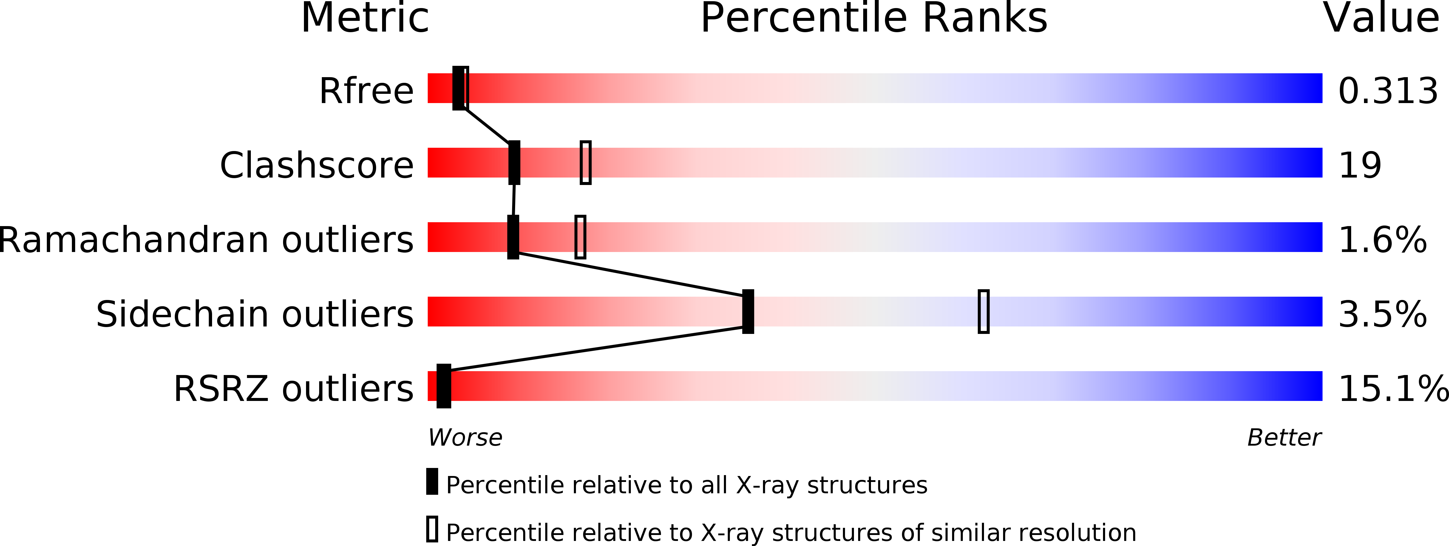
Resolution:
2.50 Å
R-Value Free:
0.30
R-Value Work:
0.28
R-Value Observed:
0.28
Space Group:
P 43