6O9B image
Deposition Date 2019-03-13
Release Date 2019-11-13
Last Version Date 2024-11-20
Entry Detail
PDB ID:
6O9B
Keywords:
Title:
Crystal structure of HLA-A3*01 in complex with a wild-type beta-catenin peptide
Biological Source:
Source Organism:
Homo sapiens (Taxon ID: 9606)
Method Details:
Experimental Method:
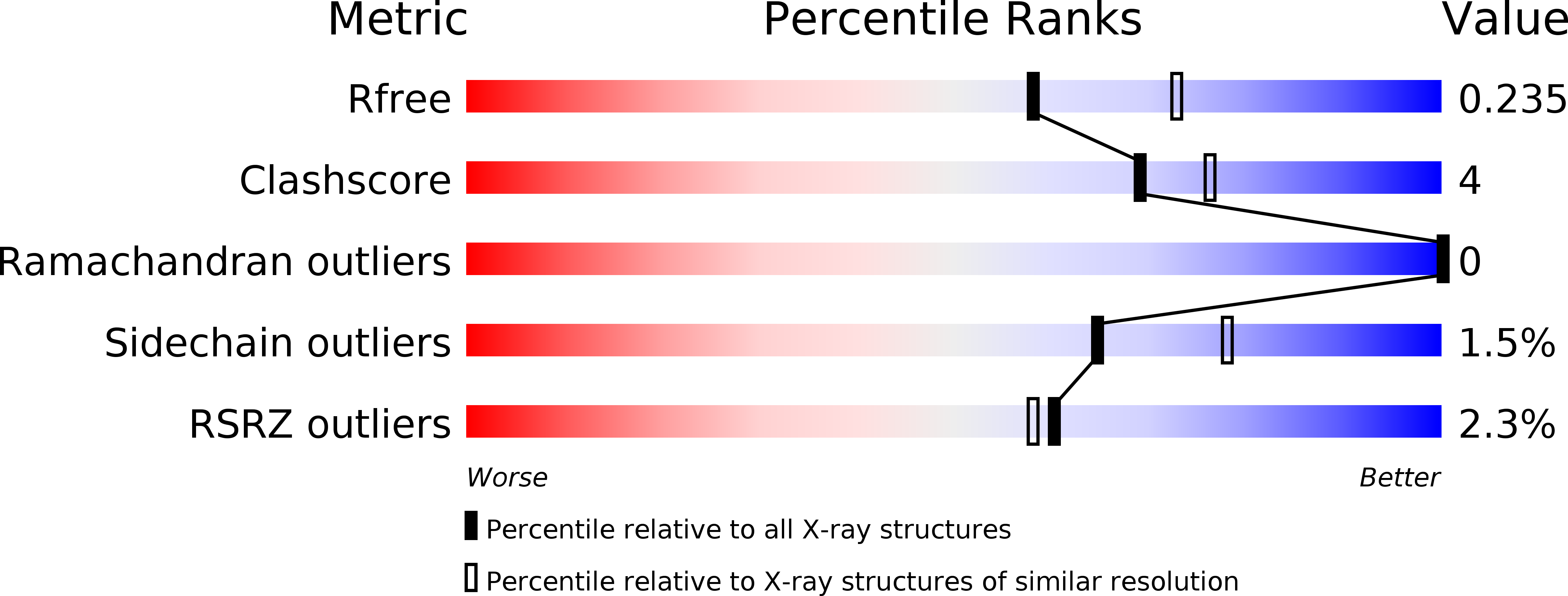
Resolution:
2.20 Å
R-Value Free:
0.22
R-Value Work:
0.18
R-Value Observed:
0.18
Space Group:
P 6 2 2
Macromolecular Entities
Polymer Type:polypeptide(L)
Molecule:HLA class I histocompatibility antigen, A-3 alpha chain
Gene (Uniprot):HLA-A
Chain IDs:A
Chain Length:300
Number of Molecules:1
Biological Source:Homo sapiens
Polymer Type:polypeptide(L)
Molecule:Beta-2-microglobulin
Gene (Uniprot):B2M
Chain IDs:B
Chain Length:119
Number of Molecules:1
Biological Source:Homo sapiens
Polymer Type:polypeptide(L)
Molecule:Catenin beta-1
Gene (Uniprot):CTNNB1
Chain IDs:C
Chain Length:9
Number of Molecules:1
Biological Source:Homo sapiens
Primary Citation
An engineered antibody fragment targeting mutant beta-catenin via major histocompatibility complex I neoantigen presentation.
J.Biol.Chem. 294 19322 19334 (2019)
PMID: 31690625 DOI: 10.1074/jbc.RA119.010251

Abstact

Mutations in CTNNB1, the gene encoding β-catenin, are common in colon and liver cancers, the most frequent mutation affecting Ser-45 in β-catenin. Peptides derived from WT β-catenin have previously been shown to be presented on the cell surface as part of major histocompatibility complex (MHC) class I, suggesting an opportunity for targeting this common driver gene mutation with antibody-based therapies. Here, crystal structures of both the WT and S45F mutant peptide bound to HLA-A*03:01 at 2.20 and 2.45 Å resolutions, respectively, confirmed the accessibility of the phenylalanine residue for antibody recognition. Phage display was then used to identify single-chain variable fragment clones that selectively bind the S45F mutant peptide presented in HLA-A*03:01 and have minimal WT or other off-target binding. Following the initial characterization of five clones, we selected a single clone, E10, for further investigation. We developed a computational model of the binding of E10 to the mutant peptide-bound HLA-A3, incorporating data from affinity maturation as initial validation. In the future, our model may be used to design clones with maintained specificity and higher affinity. Such derivatives could be adapted into either cell-based (CAR-T) or protein-based (bispecific T-cell engagers) therapies to target cancer cells harboring the S45F mutation in CTNNB1.

Legend

Protein

Chemical

Disease

Primary Citation of related structures