6O6C image
Deposition Date 2019-03-05
Release Date 2019-06-26
Last Version Date 2024-03-20
Entry Detail
PDB ID:
6O6C
Title:
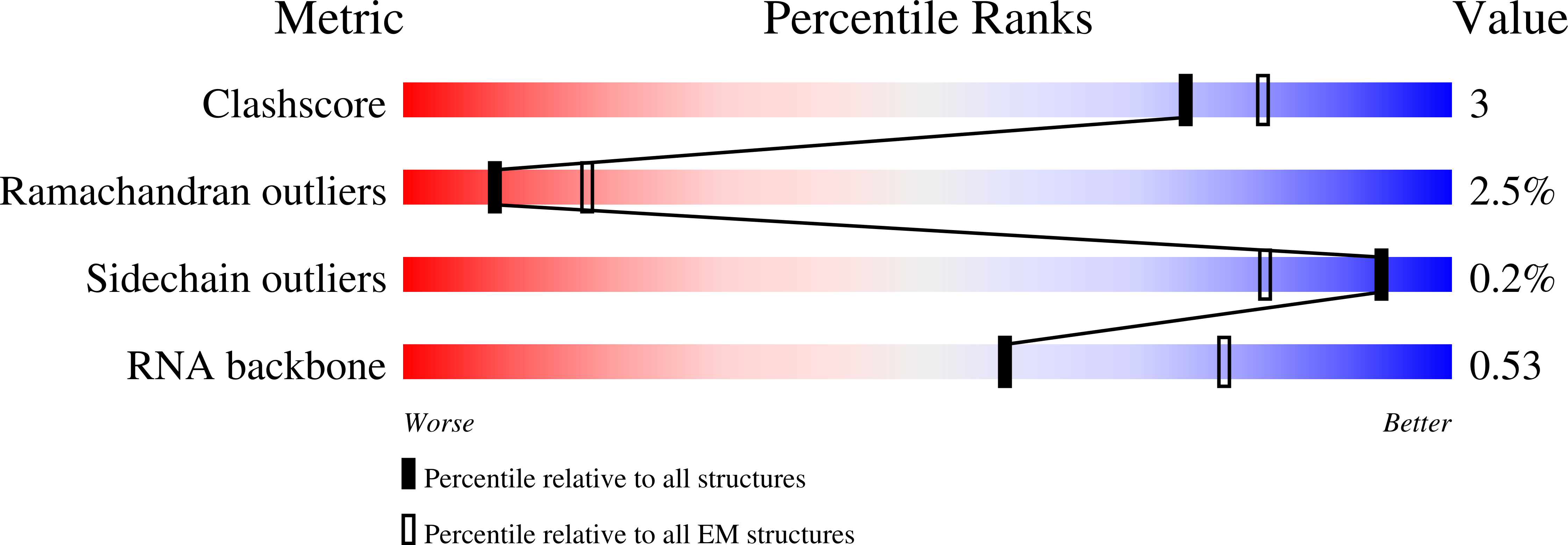
RNA polymerase II elongation complex arrested at a CPD lesion
Biological Source:
Source Organism:
Method Details:
Experimental Method:
Resolution:
3.10 Å
Aggregation State:
PARTICLE
Reconstruction Method:
SINGLE PARTICLE
Macromolecular Entities
Polymer Type:polypeptide(L)
Molecule:DNA-directed RNA polymerase II subunit RPB1
Gene (Uniprot):RPO21
Chain IDs:A
Chain Length:1733
Number of Molecules:1
Biological Source:Saccharomyces cerevisiae
Polymer Type:polypeptide(L)
Molecule:DNA-directed RNA polymerase II subunit RPB2
Gene (Uniprot):RPB2
Chain IDs:B
Chain Length:1224
Number of Molecules:1
Biological Source:Saccharomyces cerevisiae
Polymer Type:polypeptide(L)
Molecule:DNA-directed RNA polymerase II subunit RPB3
Gene (Uniprot):RPB3
Chain IDs:C
Chain Length:318
Number of Molecules:1
Biological Source:Saccharomyces cerevisiae
Polymer Type:polypeptide(L)
Molecule:DNA-directed RNA polymerases I, II, and III subunit RPABC1
Gene (Uniprot):RPB5
Chain IDs:D
Chain Length:215
Number of Molecules:1
Biological Source:Saccharomyces cerevisiae
Polymer Type:polypeptide(L)
Molecule:DNA-directed RNA polymerases I, II, and III subunit RPABC2
Gene (Uniprot):RPO26
Chain IDs:E
Chain Length:155
Number of Molecules:1
Biological Source:Saccharomyces cerevisiae
Polymer Type:polypeptide(L)
Molecule:DNA-directed RNA polymerases I, II, and III subunit RPABC3
Gene (Uniprot):RPB8
Chain IDs:F
Chain Length:146
Number of Molecules:1
Biological Source:Saccharomyces cerevisiae
Polymer Type:polypeptide(L)
Molecule:DNA-directed RNA polymerase II subunit RPB9
Gene (Uniprot):RPB9
Chain IDs:G
Chain Length:122
Number of Molecules:1
Biological Source:Saccharomyces cerevisiae
Polymer Type:polypeptide(L)
Molecule:DNA-directed RNA polymerases I, II, and III subunit RPABC5
Gene (Uniprot):RPB10
Chain IDs:H
Chain Length:70
Number of Molecules:1
Biological Source:Saccharomyces cerevisiae
Polymer Type:polypeptide(L)
Molecule:DNA-directed RNA polymerase II subunit RPB11
Gene (Uniprot):RPB11
Chain IDs:I
Chain Length:120
Number of Molecules:1
Biological Source:Saccharomyces cerevisiae
Polymer Type:polypeptide(L)
Molecule:DNA-directed RNA polymerases I, II, and III subunit RPABC4
Gene (Uniprot):RPC10
Chain IDs:J
Chain Length:70
Number of Molecules:1
Biological Source:Saccharomyces cerevisiae
Polymer Type:polyribonucleotide
Molecule:RNA (5'-R(P*AP*UP*CP*GP*AP*GP*AP*GP*G)-3')
Chain IDs:K
Chain Length:9
Number of Molecules:1
Biological Source:Saccharomyces cerevisiae
Polymer Type:polydeoxyribonucleotide
Molecule:DNA (5'-D(P*GP*GP*AP*GP*AP*AP*GP*GP*AP*GP*CP*AP*GP*AP*GP*C)-3')
Chain IDs:L
Chain Length:16
Number of Molecules:1
Biological Source:Saccharomyces cerevisiae
Polymer Type:polydeoxyribonucleotide
Molecule:DNA (27-MER)
Chain IDs:M
Chain Length:27
Number of Molecules:1
Biological Source:Saccharomyces cerevisiae
Primary Citation
3.1 angstrom structure of yeast RNA polymerase II elongation complex stalled at a cyclobutane pyrimidine dimer lesion solved using streptavidin affinity grids.
J.Struct.Biol. 207 270 278 (2019)
PMID: 31200019 DOI: 10.1016/j.jsb.2019.06.004

Abstact

Despite significant advances in all aspects of single particle cryo-electron microscopy (cryo-EM), specimen preparation still remains a challenge. During sample preparation, macromolecules interact with the air-water interface, which often leads to detrimental effects such as denaturation or adoption of preferred orientations, ultimately hindering structure determination. Randomly biotinylating the protein of interest (for example, at its primary amines) and then tethering it to a cryo-EM grid coated with two-dimensional crystals of streptavidin (acting as an affinity surface) can prevent the protein from interacting with the air-water interface. Recently, this approach was successfully used to solve a high-resolution structure of a test sample, a bacterial ribosome. However, whether this method can be used for samples where interaction with the air-water interface has been shown to be problematic remains to be determined. Here we report a 3.1 Å structure of an RNA polymerase II elongation complex stalled at a cyclobutane pyrimidine dimer lesion (Pol II EC(CPD)) solved using streptavidin grids. Our previous attempt to solve this structure using conventional sample preparation methods resulted in a poor quality cryo-EM map due to Pol II EC(CPD)'s adopting a strong preferred orientation. Imaging the same sample on streptavidin grids improved the angular distribution of its view, resulting in a high-resolution structure. This work shows that streptavidin affinity grids can be used to address known challenges posed by the interaction with the air-water interface.

Legend

Protein

Chemical

Disease

Primary Citation of related structures