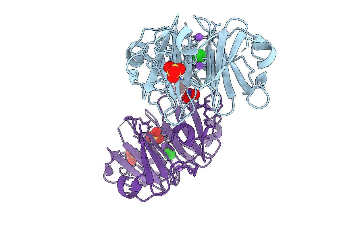
6O5E image
Deposition Date 2019-03-01
Release Date 2019-09-18
Last Version Date 2024-10-30
Entry Detail
PDB ID:
6O5E
Keywords:
Title:
Crystal structure of the Vitronectin hemopexin-like domain
Biological Source:
Source Organism:
Homo sapiens (Taxon ID: 9606)
Method Details:
Experimental Method:
Resolution:
1.90 Å
R-Value Free:
0.21
R-Value Work:
0.17
R-Value Observed:
0.18
Space Group:
P 1 21 1
Macromolecular Entities
Polymer Type:polypeptide(L)
Molecule:Vitronectin
Gene (Uniprot):VTN
Mutations:C180S, C215S
Chain IDs:A, B
Chain Length:204
Number of Molecules:2
Biological Source:Homo sapiens
Primary Citation
Structure of human Vitronectin C-terminal domain and interaction withYersinia pestisouter membrane protein Ail.
Sci Adv 5 eaax5068 eaax5068 (2019)
PMID: 31535027 DOI: 10.1126/sciadv.aax5068

Abstact

Vitronectin (Vn) is a major component of blood that controls many processes central to human biology. It is a drug target and a key factor in cell and tissue engineering applications, but despite long-standing efforts, little is known about the molecular basis for its functions. Here, we define the domain organization of Vn, report the crystal structure of its carboxyl-terminal domain, and show that it harbors the binding site for the Yersinia pestis outer membrane protein Ail, which recruits Vn to the bacterial cell surface to evade human host defenses. Vn forms a single four-bladed β/α-propeller that serves as a hub for multiple functions. The structure explains key features of native Vn and provides a blueprint for understanding and targeting this essential human protein.

Legend

Protein

Chemical

Disease

Primary Citation of related structures