6O4G image
Deposition Date 2019-02-28
Release Date 2019-07-24
Last Version Date 2023-11-15
Entry Detail
PDB ID:
6O4G
Keywords:
Title:
Structure of ALDH7A1 mutant P169S complexed with alpha-aminoadipate
Biological Source:
Source Organism:
Homo sapiens (Taxon ID: 9606)
Host Organism:
Method Details:
Experimental Method:
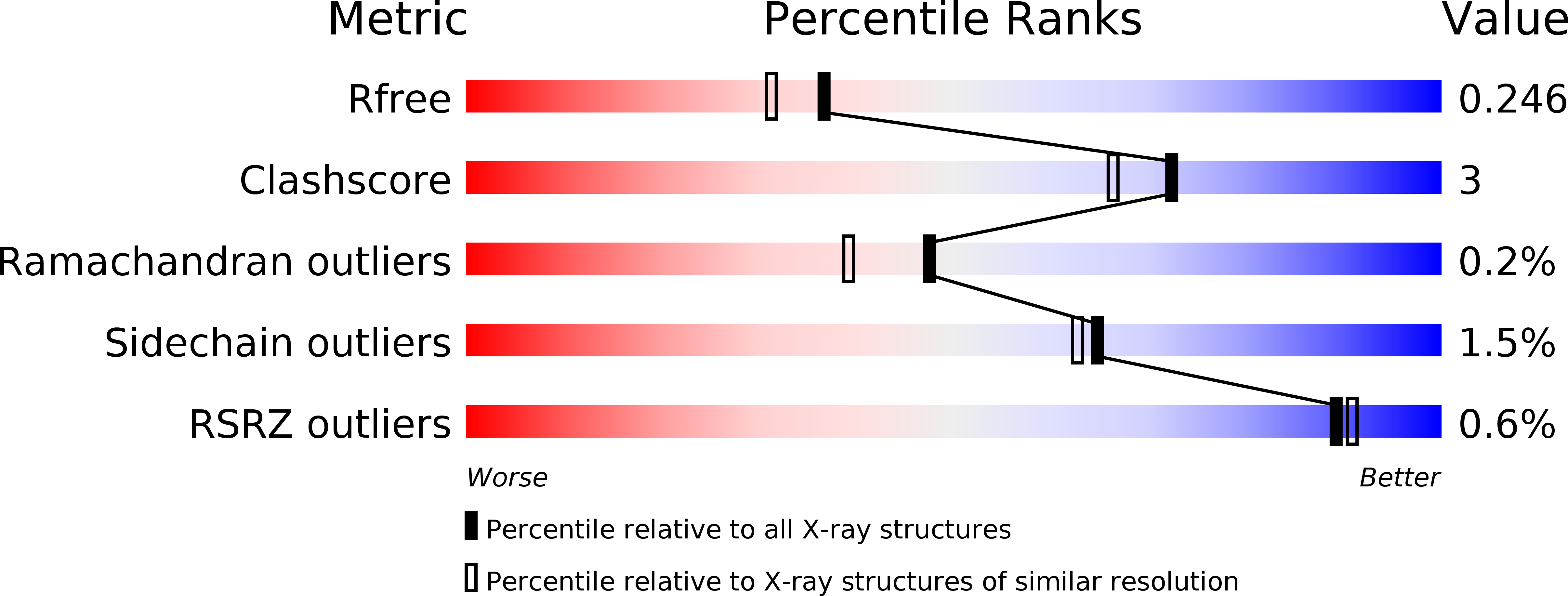
Resolution:
2.05 Å
R-Value Free:
0.25
R-Value Work:
0.19
R-Value Observed:
0.19
Space Group:
C 1 2 1
Macromolecular Entities
Polymer Type:polypeptide(L)
Molecule:Alpha-aminoadipic semialdehyde dehydrogenase
Gene (Uniprot):ALDH7A1
Mutations:P169S
Chain IDs:A, B, C, D, E, F, G, H
Chain Length:513
Number of Molecules:8
Biological Source:Homo sapiens
Primary Citation
Structural and biochemical consequences of pyridoxine-dependent epilepsy mutations that target the aldehyde binding site of aldehyde dehydrogenase ALDH7A1.
Febs J. 287 173 189 (2020)
PMID: 31302938 DOI: 10.1111/febs.14997

Abstact

In humans, certain mutations in the gene encoding aldehyde dehydrogenase 7A1 are associated with pyridoxine-dependent epilepsy (PDE). Understanding the impact of PDE-causing mutations on the structure and activity of ALDH7A1 could allow for the prediction of symptom-severity and aid the development of patient-specific medical treatments. Herein, we investigate the biochemical and structural consequences of PDE missense mutations targeting residues in the aldehyde substrate binding site: N167S, P169S, A171V, G174V, and W175G. All but G174V could be purified for biochemical and X-ray crystallographic analysis. W175G has a relatively mild kinetic defect, exhibiting a fivefold decrease in kcat with no change in Km . P169S and N167S have moderate defects, characterized by catalytic efficiencies of 20- and 100-times lower than wild-type, respectively. A171V has a profound functional defect, with catalytic efficiency 2000-times lower than wild-type. The crystal structures of the variants are the first for any PDE-associated mutant of ALDH7A1. The structures show that missense mutations that decrease the steric bulk of the side chain tend to create a cavity in the active site. The protein responds by relaxing into the vacant space, and this structural perturbation appears to cause misalignment of the aldehyde substrate in W175G and N167S. The P169S structure is nearly identical to that of the wild-type enzyme; however, analysis of B-factors suggests the catalytic defect may result from altered protein dynamics. The A171V structure suggests that the potential for steric clash with Val171 prevents Glu121 from ion pairing with the amino group of the aldehyde substrate. ENZYMES: Aldehyde dehydrogenase 7A1 (EC1.2.1.31). DATABASES: Coordinates have been deposited in the Protein Data Bank under the following accession codes: 6O4B, 6O4C, 6O4D, 6O4E, 6O4F, 6O4G, 6O4H.

Legend

Protein

Chemical

Disease

Primary Citation of related structures