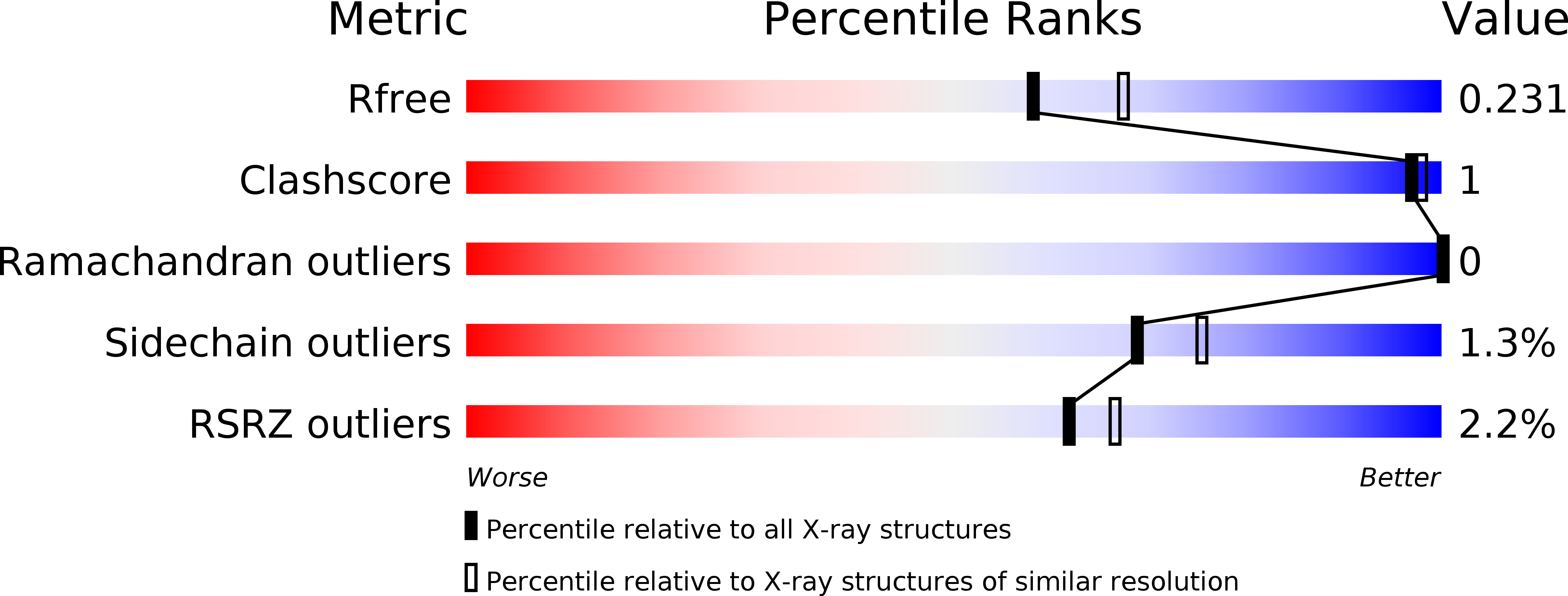
6NVQ image
Deposition Date 2019-02-05
Release Date 2019-05-22
Last Version Date 2024-05-15
Entry Detail
PDB ID:
6NVQ
Keywords:
Title:
Crystal structure of the VASH1-SVBP complex
Biological Source:
Source Organism:
Homo sapiens (Taxon ID: 9606)
Host Organism:
Method Details:
Experimental Method:
Resolution:
2.10 Å
R-Value Free:
0.21
R-Value Work:
0.19
R-Value Observed:
0.19
Space Group:
P 21 21 2
Macromolecular Entities
Polymer Type:polypeptide(L)
Molecule:Small vasohibin-binding protein
Gene (Uniprot):SVBP
Chain IDs:B
Chain Length:71
Number of Molecules:1
Biological Source:Homo sapiens
Polymer Type:polypeptide(L)
Molecule:Tubulinyl-Tyr carboxypeptidase 1
Gene (Uniprot):VASH1
Mutations:C169A
Chain IDs:A (auth: C)
Chain Length:320
Number of Molecules:1
Biological Source:Homo sapiens
Primary Citation
Crystal structure of the tubulin tyrosine carboxypeptidase complex VASH1-SVBP.
Nat.Struct.Mol.Biol. 26 567 570 (2019)
PMID: 31270470 DOI: 10.1038/s41594-019-0254-6

Abstact

The cyclic enzymatic removal and ligation of the C-terminal tyrosine of α-tubulin generates heterogeneous microtubules and affects their functions. Here we describe the crystal and solution structure of the tubulin carboxypeptidase complex between vasohibin (VASH1) and small vasohibin-binding protein (SVBP), which folds in a long helix, which stabilizes the VASH1 catalytic domain. This structure, combined with molecular docking and mutagenesis experiments, reveals which residues are responsible for recognition and cleavage of the tubulin C-terminal tyrosine.

Legend

Protein

Chemical

Disease

Primary Citation of related structures