Deposition Date
2019-01-30
Release Date
2019-07-17
Last Version Date
2024-10-23
Entry Detail
PDB ID:
6NTX
Keywords:
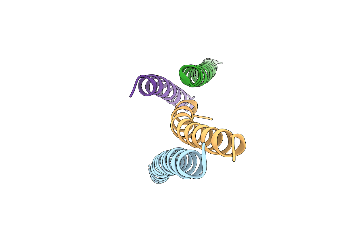
Title:
Respiratory syncytial virus fusion protein N-terminal heptad repeat domain+VIQKI
Biological Source:
Source Organism(s):
Human respiratory syncytial virus A (Taxon ID: 208893)
Human respirovirus 3 (Taxon ID: 11216)
Human respirovirus 3 (Taxon ID: 11216)
Method Details:
Experimental Method:
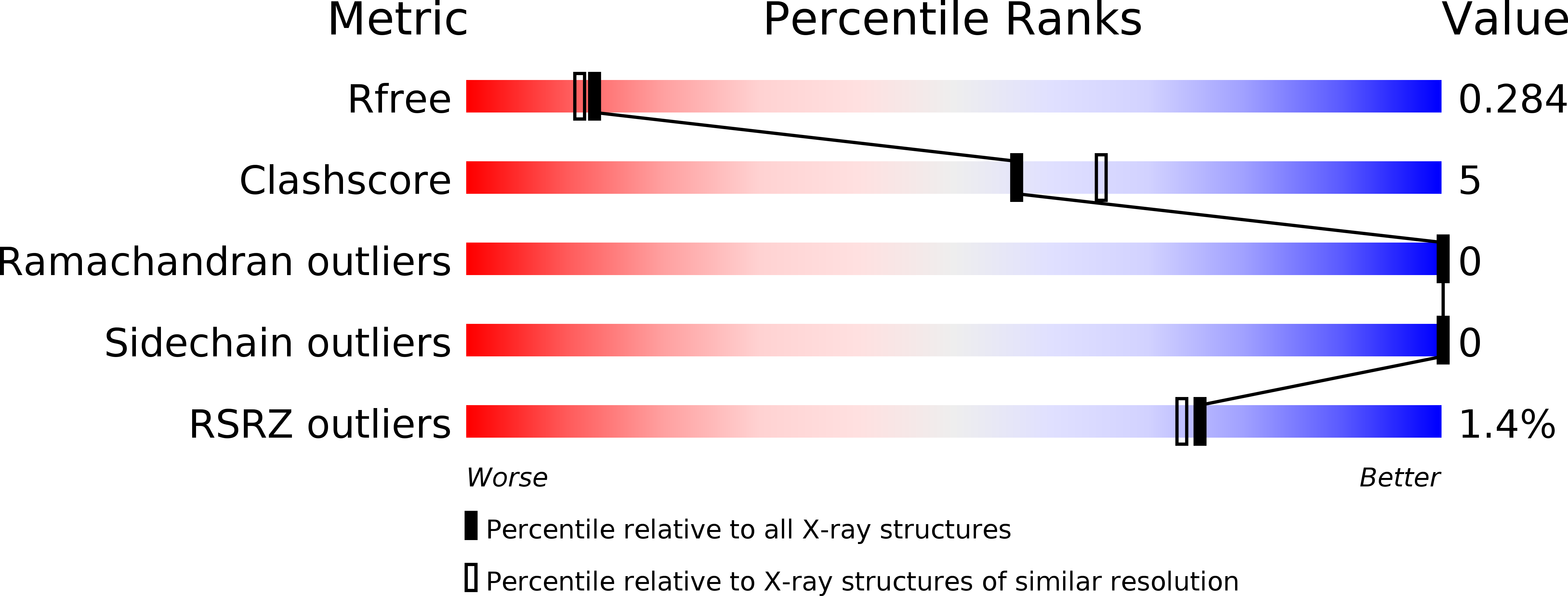
Resolution:
2.20 Å
R-Value Free:
0.28
R-Value Work:
0.23
R-Value Observed:
0.23
Space Group:
H 3 2