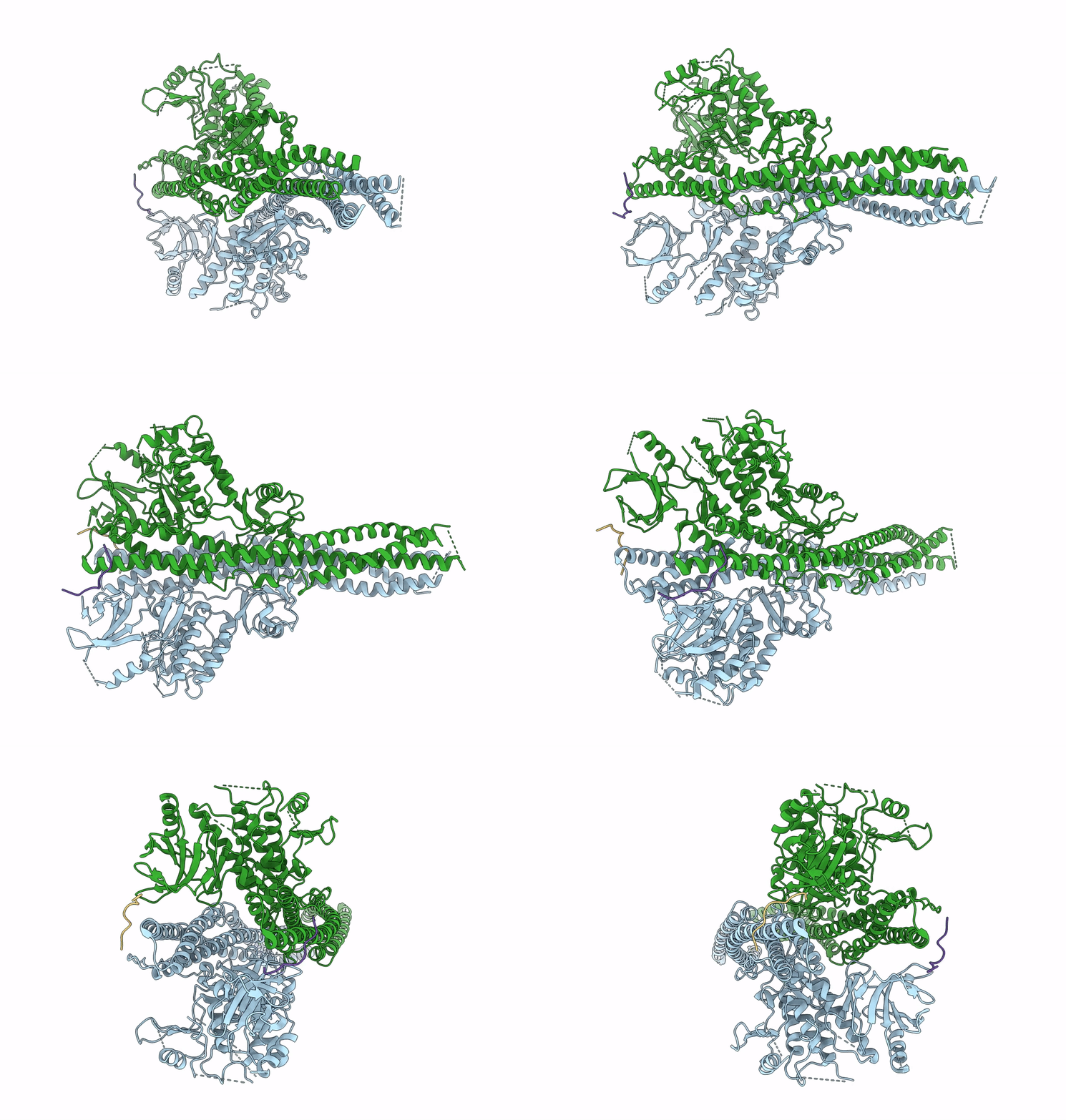
6NT9 image
Deposition Date 2019-01-28
Release Date 2019-03-06
Last Version Date 2024-03-20
Entry Detail
PDB ID:
6NT9
Keywords:
Title:
Cryo-EM structure of the complex between human TBK1 and chicken STING
Biological Source:
Source Organism:
Homo sapiens (Taxon ID: 9606)
Gallus gallus (Taxon ID: 9031)
Host Organism:
Method Details:
Experimental Method:
Resolution:
3.30 Å
Aggregation State:
PARTICLE
Reconstruction Method:
SINGLE PARTICLE
Macromolecular Entities
Structures with similar UniProt ID
Protein Blast
Polymer Type:polypeptide(L)
Molecule:Serine/threonine-protein kinase TBK1
Gene (Uniprot):TBK1
Mutagens:D135N
Chain IDs:A, C (auth: B)
Chain Length:742
Number of Molecules:2
Biological Source:Homo sapiens
Structures with similar UniProt ID
Protein Blast
Polymer Type:polypeptide(L)
Molecule:Stimulator of interferon genes protein
Chain IDs:B (auth: C), D
Chain Length:392
Number of Molecules:2
Biological Source:Gallus gallus
Ligand Molecules
Primary Citation
Structural basis of STING binding with and phosphorylation by TBK1.
Nature 567 394 398 (2019)
PMID: 30842653 DOI: 10.1038/s41586-019-1000-2

Abstact

The invasion of mammalian cytoplasm by microbial DNA from infectious pathogens or by self DNA from the nucleus or mitochondria represents a danger signal that alerts the host immune system1. Cyclic GMP-AMP synthase (cGAS) is a sensor of cytoplasmic DNA that activates the type-I interferon pathway2. On binding to DNA, cGAS is activated to catalyse the synthesis of cyclic GMP-AMP (cGAMP) from GTP and ATP3. cGAMP functions as a second messenger that binds to and activates stimulator of interferon genes (STING)3-9. STING then recruits and activates tank-binding kinase 1 (TBK1), which phosphorylates STING and the transcription factor IRF3 to induce type-I interferons and other cytokines10,11. However, how cGAMP-bound STING activates TBK1 and IRF3 is not understood. Here we present the cryo-electron microscopy structure of human TBK1 in complex with cGAMP-bound, full-length chicken STING. The structure reveals that the C-terminal tail of STING adopts a β-strand-like conformation and inserts into a groove between the kinase domain of one TBK1 subunit and the scaffold and dimerization domain of the second subunit in the TBK1 dimer. In this binding mode, the phosphorylation site Ser366 in the STING tail cannot reach the kinase-domain active site of bound TBK1, which suggests that STING phosphorylation by TBK1 requires the oligomerization of both proteins. Mutational analyses validate the interaction mode between TBK1 and STING and support a model in which high-order oligomerization of STING and TBK1, induced by cGAMP, leads to STING phosphorylation by TBK1.

Legend

Protein

Chemical

Disease

Primary Citation of related structures