6NQR image
Entry Detail
PDB ID:
6NQR
Title:
Crystal structure of fast switching M159T mutant of fluorescent protein Dronpa (Dronpa2)- Y63(3-NO2Y)
Biological Source:
Source Organism:
PDB Version:
Deposition Date:
2019-01-21
Release Date:
2019-06-12
Method Details:
Experimental Method:
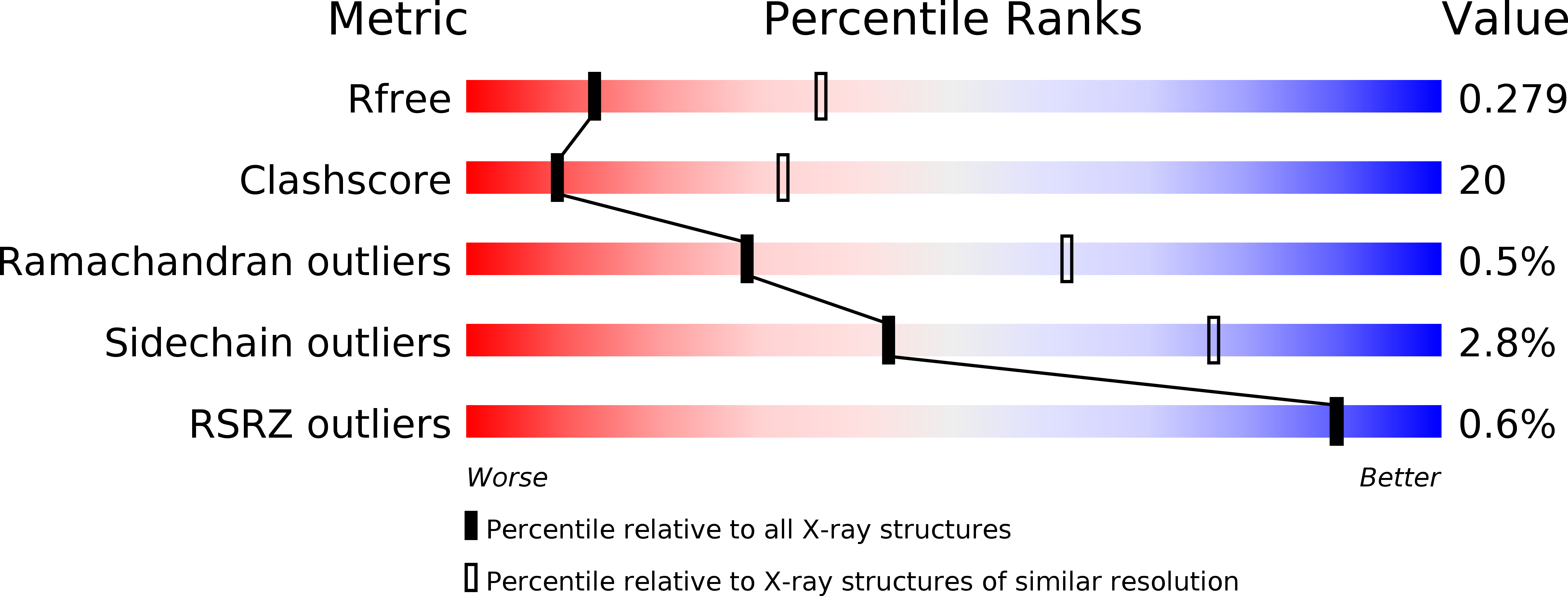
Resolution:
2.90 Å
R-Value Free:
0.28
R-Value Work:
0.21
R-Value Observed:
0.22
Space Group:
P 1
Macromolecular Entities
Polymer Type:polypeptide(L)
Description:Fluorescent protein Dronpa
Mutations:M159T, E218G
Chain IDs:A, B, C, D, E, F, G, H
Chain Length:255
Number of Molecules:8
Biological Source:Echinophyllia sp. SC22
Modified Residue
Compound ID Chain ID Parent Comp ID Details 2D Image
KY7 A CYS chromophore
Ligand Molecules
Primary Citation
Electrostatic control of photoisomerization pathways in proteins.
Science 367 76 79 (2020)
PMID: 31896714 DOI: 10.1126/science.aax1898

Abstact

Rotation around a specific bond after photoexcitation is central to vision and emerging opportunities in optogenetics, super-resolution microscopy, and photoactive molecular devices. Competing roles for steric and electrostatic effects that govern bond-specific photoisomerization have been widely discussed, the latter originating from chromophore charge transfer upon excitation. We systematically altered the electrostatic properties of the green fluorescent protein chromophore in a photoswitchable variant, Dronpa2, using amber suppression to introduce electron-donating and electron-withdrawing groups to the phenolate ring. Through analysis of the absorption (color), fluorescence quantum yield, and energy barriers to ground- and excited-state isomerization, we evaluate the contributions of sterics and electrostatics quantitatively and demonstrate how electrostatic effects bias the pathway of chromophore photoisomerization, leading to a generalized framework to guide protein design.

Legend

Protein

Chemical

Disease

Primary Citation of related structures